| CPC G16H 50/50 (2018.01) [G06N 3/045 (2023.01); G06N 3/047 (2023.01); G06N 3/048 (2023.01); G06N 3/088 (2013.01); G06T 7/11 (2017.01); G16H 30/40 (2018.01); G16H 50/20 (2018.01); G06T 2207/10081 (2013.01); G06T 2207/10088 (2013.01); G06T 2207/20081 (2013.01); G06T 2207/20084 (2013.01); G06T 2207/30048 (2013.01); G06T 2207/30056 (2013.01); G06T 2207/30064 (2013.01)] | 36 Claims |

|
1. A machine learning system, comprising:
one or more nontransitory processor-readable storage media that collectively store at least one of processor-executable instructions or data; and
one or more processors communicably coupled to the one or more nontransitory processor-readable storage media, wherein in operation, the one or more processors individually or collectively:
receives learning data comprising a plurality of batches of labeled image sets and corresponding segmentation masks, each image set comprising image data representative of an input anatomical structure;
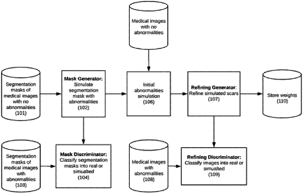
simulates abnormalities on at least some images in the image sets and their corresponding segmentation masks without abnormalities, using a plurality of trained convolutional neural network (CNN) models stored on the at least one nontransitory processor-readable storage medium, wherein the abnormalities are in at least one of (a) cardiac images, and include one or more of myocardial scarring, myocardial infarction, coronary stenosis, or perfusion characteristics of the cardiac myocardium, (b) lung images, and include one or more of ground glass, solid lung nodules, or sub-solid lung nodules, or (c) liver images, and include one or more of Hemangiomas, Focal Nodular Hyperplasias lesions, Hepatocellualar Adenomas lesions, or cystic masses; and
stores the simulated abnormalities as images and corresponding segmentation masks in the at least one nontransitory processor-readable storage medium of the machine learning system.
|