| CPC A61B 8/4263 (2013.01) [A61B 8/4245 (2013.01); A61B 8/463 (2013.01); A61B 8/5223 (2013.01); G06T 7/0012 (2013.01); G06T 7/70 (2017.01); G06T 11/00 (2013.01); G06V 10/17 (2022.01); G06T 2207/10016 (2013.01); G06T 2207/10132 (2013.01); G06T 2207/30061 (2013.01)] | 22 Claims |
|
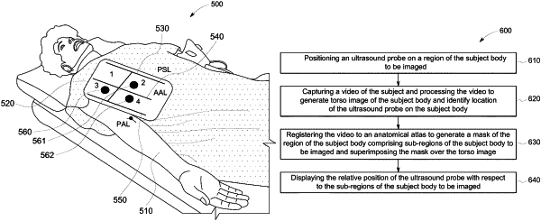
1. A method for guided coverage and automated detection of pathologies of a subject body, the method comprising:
positioning an ultrasound probe on a subject's torso;
capturing a video of the subject body and processing the video to generate a torso image of the subject body and identify a location of the ultrasound probe with respect to the subject body;
registering the video to an anatomical atlas to generate a mask of a region of the subject body comprising a plurality of sub-regions of the subject body to be imaged and superimposing the mask over the torso image;
displaying an indicator corresponding to a location of the each of the plurality of the sub-regions on the torso image; and
displaying a relative position of the ultrasound probe with respect to the indicator corresponding to the location of the each of the plurality of sub-regions of the subject body to be imaged.
|
|
16. A system for guided coverage and automated detection of pathologies of a subject body, the system comprising:
a portable device comprising a camera and a display configured to acquire a video stream of the subject body;
an ultrasound probe positioned over a subject's torso and connected to the portable device; and
a computer system connected to the portable device and configured to receive a plurality of ultrasound images and a video stream of the subject body, wherein the computer system comprises:
a processor;
a memory connected to the processor;
at least one artificial intelligence module deployed over the memory and configured to generate a torso image of the subject body;
an atlas module deployed over the memory and configured generate a mask of a region of the subject body comprising a plurality of sub-regions to be imaged and superimpose the mask over the torso image of the subject body;
wherein the computer system is further configured to display an indicator corresponding to a location of the each of the plurality of the sub-regions on the torso image, and display a relative position of the ultrasound probe with respect to the indicator corresponding to the location of the each of the plurality of sub-regions of the subject body.
|