| CPC G06V 20/698 (2022.01) [G06T 7/0014 (2013.01); G06T 7/74 (2017.01); G06T 7/90 (2017.01); G06V 10/44 (2022.01); G06V 10/56 (2022.01); G06T 2207/20084 (2013.01); G06T 2207/30024 (2013.01)] | 14 Claims |

|
1. An image analysis method for analyzing an image of a tissue or a cell to be analyzed using a deep learning algorithm of a neural network structure, the method comprising:
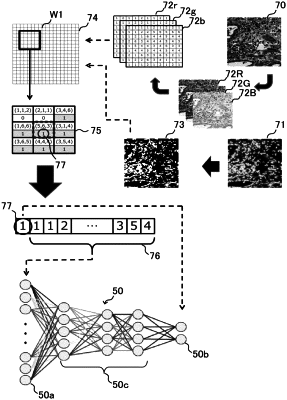
generating analysis data from an analysis target image that include the tissue or the cell to be analyzed;
inputting the analysis data to the deep learning algorithm, and
generating data indicating a region of a cell nucleus in the analysis target image by the deep learning algorithm,
wherein training data used for learning of the deep learning algorithm are generated based on a bright field image of a tissue specimen or a sample containing a cell captured under a bright field microscope and a fluorescence image of a cell nucleus in the same tissue specimen or the same sample prepared by applying fluorescent nuclear stain to the same tissue specimen or the same sample for fluorescence observation by a fluorescence microscope, wherein the fluorescence image is converted to binary data indicating which part of the fluorescence image is the cell nucleus region, and
the analysis target image is a bright field image.
|