| CPC A61B 6/487 (2013.01) [A61B 6/461 (2013.01); A61B 6/481 (2013.01); A61B 6/504 (2013.01); A61B 6/5211 (2013.01); A61B 6/5294 (2013.01)] | 11 Claims |

|
1. An X-ray fluoroscopic imaging apparatus comprising:
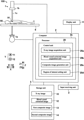
an imaging unit including an X-ray source for irradiating an object with X-rays and an X-ray detector for detecting X-rays emitted from the X-ray source;
an X-ray image acquisition unit configured to acquire an X-ray image captured by the imaging unit;
a blood vessel extracted image acquisition unit configured to acquire a blood vessel extracted image in which a blood vessel image of the object is extracted, the blood vessel image having been generated in advance based on a contrast image that is the X-ray image captured with a contrast agent administered to the object;
a composite image generation unit configured to generate a first composite image in which the X-ray image captured with no contrast agent administered and the blood vessel extracted image are composed with the X-ray image and the blood vessel extracted image aligned in positions; and
a region of interest setting unit configured to set a region of interest, the region of interest being a part of the X-ray image and reflecting a device introduced into a blood vessel of the object,
wherein the composite image generation unit is configured to generate a second composite image by aligning positions of the X-ray image and the blood vessel extracted image, based on the device reflected in the region of interest set by the region of interest setting unit and the blood vessel image reflected in the blood vessel extracted image.
|