| CPC A61B 5/0068 (2013.01) [A61B 5/7267 (2013.01); A61B 90/361 (2016.02); A61B 90/37 (2016.02); G06N 3/045 (2023.01); G06N 3/08 (2013.01); G06T 7/0012 (2013.01); G06V 10/454 (2022.01); G06V 10/764 (2022.01); G06V 10/82 (2022.01); G06V 20/693 (2022.01); G06V 20/698 (2022.01); G16H 30/20 (2018.01); G16H 30/40 (2018.01); G16H 50/20 (2018.01); A61B 2090/373 (2016.02); G06T 2207/20084 (2013.01); G06T 2207/30016 (2013.01)] | 20 Claims |
|
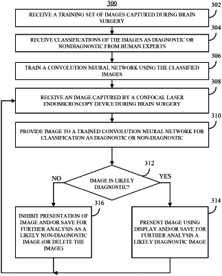
1. A method for selectively presenting images captured by a confocal laser endomicroscopy (CLE) device, comprising:
receiving a first plurality of images captured by a CLE device during brain surgery;
providing each of the first plurality of images to a convolutional neural network (CNN) trained using at least a second plurality of images, wherein each of the second plurality of images is an image of brain tissue that was captured using CLE techniques and is labeled as either a diagnostic image or a non-diagnostic image, wherein images labeled as diagnostic provide at least a threshold level of identifiable histological features and images labeled as non-diagnostic do not provide the threshold level of identifiable histological features;
identifying, based on outputs of the CNN, a first subset of the first plurality of images as non-diagnostic images; and
identifying, based on outputs of the CNN, a second subset of the first plurality of images as diagnostic images.
|