| CPC G06T 7/0012 (2013.01) [G06T 7/12 (2017.01); G16H 15/00 (2018.01); G06T 5/30 (2013.01); G06T 2207/10024 (2013.01); G06T 2207/20081 (2013.01); G06T 2207/20084 (2013.01); G06T 2207/30041 (2013.01)] | 2 Claims |

|
1. A three-way U-Net method for accurately segmenting an uncertain boundary of a retinal blood vessel, comprising the following steps:
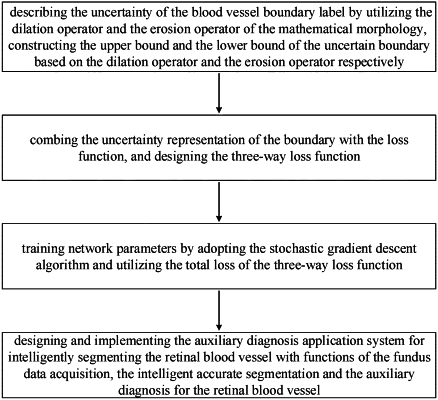
S1, constructing, in a feature encoding part of a U-net, a feature extraction module for extracting retinal vessel features layer by layer, wherein the feature extraction module includes two 3×3 convolutional layers, one Dropout layer, one Relu excitation layer, and one 2×2 maximum pooling layer; constructing, in a feature decoding part of a U-net module, a recovery feature module for recovering a size of an image and restore position information on the image, wherein the recovery feature module includes one 2×2 upsampling layer and two 3×3 convolutional layers; connecting symmetrical layers of an upsampling and a sub-sampling by a skip connection; splicing a higher layer feature map with a bottom layer feature map to serve as an input for a subsequent layer to enable the model to fuse higher features with lower features to learn more blood vessel features and obtain a more accurate output feature map; and convolving, after recovering a feature size of the image, two 1×1 convolutional kernels to obtain a feature map with a channel number of 2, wherein one channel represents a vascular category, and another channel represents a non-vascular category; and calculating, by a softmax layer, a probability that each pixel point in the image belongs to each category;
S2, describing, based on a dilation operator and an erosion operator of a mathematical morphology, an uncertainty of a retinal blood vessel boundary label; constructing, based on the dilation operator, an upper bound map Idilate of an uncertain boundary of the retinal blood vessel, and constructing, based on the erosion operator, a lower bound map Ierose of the uncertain boundary of the retinal blood vessel respectively, to obtain a maximum value and a minimum value for a blood vessel boundary; and mapping a boundary with uncertain information into one range;
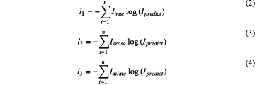
S3, combining the uncertainty of the boundary with a loss function and forming a three-way loss function; calculating, by the loss function, a cross entropy loss between a retinal blood vessel prediction boundary map Ipredict and a retinal blood vessel manual segmentation gold standard map Itrue, a cross entropy loss between the upper bound map Idilate of the uncertain boundary of the retinal blood vessel and the lower bound map Ierose of the uncertain boundary of the retinal blood vessel; and generating, by a weighted fusion mode, an eventual loss, wherein a formula for calculating the eventual loss is as follows:
 wherein l1 denotes the cross entropy loss between the retinal blood vessel prediction image Ipredict and the retinal blood vessel manual segmentation gold standard map Itrue, and l1 is calculated as shown in Formula (2); l2 denotes a cross entropy loss between the retinal blood vessel prediction image Ipredict and the lower bound map Idilate of the uncertain boundary of the retinal blood vessel constructed based on the erosion operator, l2 is calculated as shown in Formula (3); l3 denotes a cross entropy loss between the retinal blood vessel prediction image Ipredict and the upper bound map Ierose of the uncertain boundary of the retinal blood vessel constructed based on the dilation operator, l2 is calculated as shown in Formula (4); α and 1-α are hyper-parameters, α denotes a loss weight between the retinal blood vessel prediction image Ipredict and the lower bound map Ierose of the uncertain boundary of the retinal blood vessel, and 1-α denotes a loss weight between the retinal blood vessel prediction image Ipredict and the upper bound map Idilate of the uncertain boundary of the retinal blood vessel;
 wherein n denotes a number of categories contained in the images; and training by adopting a stochastic gradient descent algorithm, network parameters by utilizing a total loss of the three-way loss function; and
S4, implementing, by utilizing a Django framework in Python, an auxiliary diagnosis application system for intelligently segmenting the retinal blood vessel, wherein the application system includes a fundus data acquisition, an intelligent accurate segmentation for a retinal blood vessel and an auxiliary diagnosis for the retinal blood vessel, the fundus data acquisition is collecting basic information on a patient as well as left and right eye images of the patient captured by medical equipment, the intelligent accurate segmentation for the retinal blood vessel is segmenting the blood vessels in the captured left and right eye images of the patient by utilizing a U-Net model based on the three-way loss function (TWD-UNet), and uploading segmentation results to a remote diagnostic module; the auxiliary diagnosis is the doctor analyzes a patient's condition and provides diagnostic advice according to the basic information on the patient, color left and right eye images captured by a fundus camera and a corresponding segmented image of the retinal blood vessel to generate a downloadable and printable electronic diagnosis report
wherein the downloadable and printable electronic diagnosis report provides personalized medical services for the patient.
|