| CPC A61F 9/00827 (2013.01) [A61F 9/0084 (2013.01); A61F 2009/00872 (2013.01)] | 20 Claims |

|
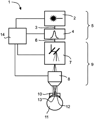
1. A device for generating a cut in an interior of an eye, the device comprising:
a contact lens comprising a contact surface for placing on a cornea of the eye;
a laser beam source designed to emit laser radiation having a pulse frequency of at least 1.2 MHz and a wavelength which penetrates the cornea, wherein the energy of each individual pulse separating tissue within the cornea having a pulse energy of 10 nJ to 80 nJ;
beam optics designed to focus the laser radiation through the contact lens into a focus located in the eye;
a beam scanner designed to shift the focus in the eye along a path within the cornea to generate the cut as a cut which confines a lenticule within the cornea, wherein the cut is, at least in parts, curved against a frontside or backside of the cornea; and
a processor, which is connected with the beam scanner and is designed to control the beam scanner by specifying the path and thus defining location and extension of the cut confining the lenticule within the cornea;
wherein the laser beam source is configured to minimize tissue-splitting separation of collagen structures of the corneal tissue, thereby improving contour accuracy of the curved cut surface and of dimensions of the isolated lenticule.
|
|
13. A method for generating a cut in an interior of an eye, the method comprising:
providing a contact lens comprising a contact surface and placing the contact surface on the cornea;
emitting laser radiation pulses having a pulse frequency of at least 1.2 MHz and a wavelength which penetrates a cornea of the eye;
focusing the laser radiation pulses into a focus located in the eye by using beam optics; and
shifting the focus in the eye along a path which defines location and extension of the cut as a cut which is within the cornea and confines a lenticule within the cornea, wherein the cut is, at least in parts, curved against a frontside or backside of the cornea;
wherein the energy of each individual pulse separating tissue within the cornea has a pulse energy of 10 nJ to 80 nJ or the beam optics comprises an objective with a numerical aperture of at least 0.33, and
wherein the laser beam source and the beam optics are configured to minimize tissue-splitting separation of collagen structures of the corneal tissue, thereby improving contour accuracy of the curved cut surface and of dimensions of the isolated lenticule.
|
|
20. A device for generating a cut in the interior of an eye, the device comprising:
a contact lens comprising a contact surface for placing on a cornea of the eye;
a laser beam source designed to emit laser radiation having a pulse frequency of at least 1.2 MHz and a wavelength which penetrates the cornea;
beam optics designed to focus the laser radiation through the contact lens into a focus located in the eye and comprising an objective with a numerical aperture of at least 0.33;
a beam scanner designed to shift the focus in the eye along a path within the cornea to generate the cut as a cut which confines a lenticule within the cornea, wherein the cut is—at least in parts—curved against a frontside or backside of the cornea; and
a processor, which is connected with the beam scanner and is designed to control the beam scanner by specifying the path and thus defining location and extension of the cut confining the lenticule within the cornea;
wherein the beam optics are configured to minimize tissue-splitting separation of collagen structures of the corneal tissue, thereby improving contour accuracy of the curved cut surface and of dimensions of the isolated lenticule.
|