| CPC A61B 34/10 (2016.02) [A61B 2018/00351 (2013.01); A61B 2034/101 (2016.02); A61B 2034/104 (2016.02); A61B 2034/105 (2016.02)] | 19 Claims |
|
1. A method for identifying one or more ablation locations in an atrial tissue region in an atrial fibrillation (AF) patient with atrial fibrosis, the method comprising:
receiving three-dimensional imaging data representing the atria of the patient;
generating a patient-specific atrial model of the atria from the three-dimensional imaging data;
conducting simulation of AF using the patient-specific atrial model to identify AF-perpetrating regions; and
identifying from the AF-perpetrating regions one or more locations in the atria suitable for guided catheter ablation to make the atria non-inducible to AF,
wherein generating the patient-specific atrial model comprises:
constructing a geometric model of at least the left atrium and the right atrium of the AF patient's heart, said geometric model including normal tissue regions and including remodeled tissue regions, wherein tissue regions are segmented into a plurality of different regions based on the three-dimensional imaging data;
retrieving reference fiber orientations from an atlas database; and
mapping the retrieved reference fiber orientations to the constructed geometric model; and
wherein the conducting the simulation comprises:
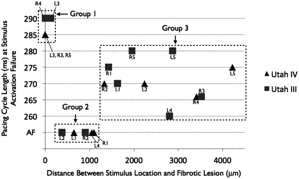
identifying multiple groups of pacing locations according to pacing cycle length and proximity to at least one lesion in the patient-specific atrial model; and
identifying at least one group from among the multiple groups that sustains AF during pacing.
|