| CPC A61B 8/445 (2013.01) [A61B 8/0883 (2013.01); A61B 8/12 (2013.01); A61B 8/4488 (2013.01); A61B 8/5207 (2013.01); A61B 90/39 (2016.02); B06B 1/0215 (2013.01); B06B 1/0622 (2013.01); A61B 2090/3966 (2016.02); B06B 2201/76 (2013.01)] | 20 Claims |

|
1. An ultrasonic imaging system comprising:
an ultrasonic catheter having a longitudinal axis, a proximal end, and a distal end;
an ultrasonic transducer array disposed within the distal end of the ultrasonic catheter,
wherein the ultrasonic transducer array comprises a substrate and a plurality of transducer array elements arranged on the substrate; and
a catheter shaft connected at one end to a handle assembly and at other end to the ultrasonic transducer array, wherein the catheter shaft houses an electronic flex cable which is in communication with at least one signal trace, and is configured to:
direct each of the plurality of transducer array elements, via the at least one signal trace, to transmit and receive, with respect to heart, ultrasound beams;
receive at least one signal from the plurality of transducer array elements based on transmitting and receiving at least one ultrasound beam of the ultrasound beams; and
construct at least one image of at least a portion of the heart based on the at least one signal,
wherein the ultrasonic catheter is coupled to an imaging device using a dongle, and the dongle is configured to communicate ultrasound transmit pulses and ultrasound receive waveforms between the imaging device and the ultrasonic catheter, wherein:
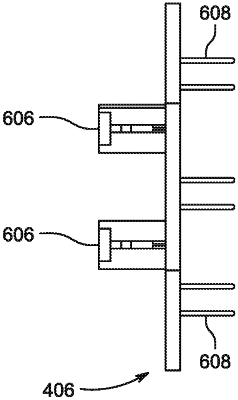
the dongle comprises an interposer located at a proximal end of the handle assembly, the interposer having a plurality of connector pins extending from a proximal side and a board edge connector extending from a distal side; and
the board edge connector is attached to a flat circuit board of the handle assembly that is connected to a plurality of individual electronic flex cables.
|
|
10. An ultrasonic catheter comprising:
a body having a longitudinal axis and a distal end;
an ultrasonic transducer array disposed within the distal end of the body, wherein the ultrasonic transducer array comprises a plurality of transducer array elements arranged on a substrate,
wherein each transducer array element comprises a plurality of transducers, with a first group of two or more transducers in a first transducer array element and a second group of two or more transducers in the first transducer array element, and each transducer array element is connected in parallel, and comprising:
at least one piezoelectric layer disposed on the substrate; at least one first electrode connected between the at least one piezoelectric layer and a signal conductor; and
at least one second electrode connected between the at least one piezoelectric layer and a ground conductor; and
wherein the ultrasonic transducer is configured to be coupled to an imaging device using a dongle, and the dongle is configured to communicate ultrasound transmit pulses and ultrasound receive waveforms between the imaging device and the ultrasonic catheter, wherein:
the dongle comprises an interposer located at a proximal end of the handle assembly, the interposer having a plurality of connector pins extending from a proximal side and a board edge connector extending from a distal side; and
the board edge connector is attached to a flat circuit board of the handle assembly that is connected to a plurality of individual electronic flex cables.
|
|
19. An intracardiac echocardiographic (ICE) imaging system comprising:
an ultrasonic catheter having a longitudinal axis, a proximal end, and a distal end;
a micro-electromechanical (MEMS) based Piezoelectric Micromachined Ultrasonic Transducer (pMUT) array disposed within the distal end of the ultrasonic catheter,
wherein the MEMS based pMUT array comprises a substrate and a plurality of MEMS based pMUT array elements arranged on the substrate; and
an electronic flex cable connected at one end to a handle assembly and at other end to the MEMS based pMUT array, wherein the electronic flex cable is in communication with at least one signal trace, and is configured to:
direct each of the plurality of MEMS based pMUT array elements, via the at least one signal trace, to transmit and receive, with respect to heart, ultrasound beams;
receive at least one signal from the plurality of MEMS based pMUT array elements based on transmitting and receiving at least one ultrasound beam of the ultrasound beams; and
construct at least one image of at least a portion of the heart based on the at least one signal;
wherein the ultrasonic catheter is coupled to an imaging device using a dongle, and the dongle is configured to communicate ultrasound transmit pulses and ultrasound receive waveforms between the imaging device and the ultrasonic catheter, wherein:
the dongle comprises an interposer located at a proximal end of the handle assembly, the interposer having a plurality of connector pins extending from a proximal side and a board edge connector extending from a distal side; and
the board edge connector is attached to a flat circuit board of the handle assembly that is connected to a plurality of individual electronic flex cables.
|