| CPC A61B 6/5217 (2013.01) [A61B 5/02007 (2013.01); A61B 5/02028 (2013.01); A61B 5/4839 (2013.01); A61B 5/7275 (2013.01); A61B 6/481 (2013.01); A61B 6/504 (2013.01)] | 1 Claim |

|
1. A method for assessing coronary microvascular dysfunction of a patient, the method comprising:
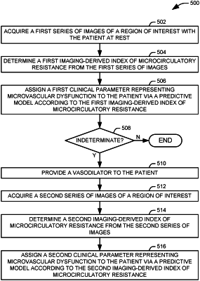
acquiring a first series of images of a region of interest within the patient;
determining a first angiography-derived index of microcirculatory resistance from the series of images and a measured first aortic pressure of the patient, wherein determining the imaging-derived index of microcirculatory resistance from the series of images comprises:
measuring the first aortic pressure of the patient;
determining an angiography-derived fractional flow reserve from the series of images;
determining, from the series of images, a transit time of a marker to travel between first and second reference points; and
calculating a product of a ratio of the number of frames necessary for the contrast dye to travel from the guiding catheter to the distal reference to a frame rate of the imager, the first aortic pressure, and the angiography-derived fractional flow reserve determined from the series of images;
assigning a first clinical parameter to the patient via a first predictive model trained on a set of training data representing the first clinical parameter and receiving at least the first angiography-derived index of microcirculatory resistance as an input and providing the first clinical parameter as an output, wherein the first clinical parameter is a categorical parameter that can assume a first value representing a presence of microvascular dysfunction, a second value representing an absence of microvascular dysfunction, and a third value representing an indeterminate result; and
performing the following steps if the first clinical parameter assumes the third value:
providing a vasodilator to the patient;
acquiring a second series of images of the region of interest;
determining a second angiography-derived index of microcirculatory resistance from the second series of images and a second measured aortic pressure of the patient; and
assigning a second clinical parameter to the patient via a second predictive model trained on a set of training data representing the second clinical parameter and receiving at least the second angiography-derived index of microcirculatory resistance as an input and providing the second clinical parameter as an output, wherein the second clinical parameter is a categorical parameter that can assume a first value representing the presence of microvascular dysfunction and a second value representing the absence of microvascular dysfunction.
|