| CPC A61B 34/37 (2016.02) [A61B 1/0005 (2013.01); A61B 1/000094 (2022.02); A61B 1/000096 (2022.02); A61B 1/00149 (2013.01); A61B 1/313 (2013.01); A61B 17/02 (2013.01); A61B 34/20 (2016.02); A61B 34/74 (2016.02); A61B 90/37 (2016.02); G06T 7/70 (2017.01); G06T 7/73 (2017.01); A61B 34/10 (2016.02); A61B 34/25 (2016.02); A61B 90/03 (2016.02); A61B 2034/2057 (2016.02); A61B 2034/2065 (2016.02); A61B 2034/302 (2016.02); A61B 2090/306 (2016.02); A61B 2090/309 (2016.02); A61B 2090/371 (2016.02); G06T 2207/10021 (2013.01); G06T 2207/30004 (2013.01)] | 19 Claims |
|
1. A method of assisting positioning of an instrument in a body cavity of a patient using a robotic surgery system, the robotic surgery system being controlled by a processor, the method comprising, by the processor:
receiving body cavity image data representing an interior view of the body cavity, the body cavity image data being captured by a camera inserted into the body cavity, the camera being detached from the robotic surgery system to facilitate positioning of the camera by hand to receive a desired interior view of the body cavity, and a position of the camera being adjusted responsive to connecting the camera to the robotic surgery system;
generating one or more display signals being configured to display a view of the interior view of the body cavity, the view being at least partially based on the body cavity image data;
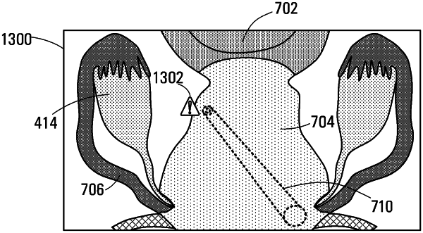
determining an instrument envelope identifying a workable volume of the instrument, the workable volume comprising a range of motion of the instrument within the body cavity;
generating an envelope overlay image, the envelope overlay image being configured to represent the instrument envelope; and
causing a display to display the view and the envelope overlay image.
|