| CPC A61B 18/1492 (2013.01) [A61B 1/00082 (2013.01); A61B 1/00096 (2013.01); A61B 1/05 (2013.01); A61B 1/0661 (2013.01); A61B 1/3132 (2013.01); A61B 2017/00243 (2013.01); A61B 2018/00392 (2013.01); A61B 2018/00577 (2013.01); A61B 2018/00791 (2013.01)] | 4 Claims |

|
1. A dual-probe medical device, comprising:
a sheath;
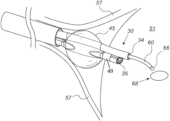
a first probe, said first probe being a pericardium catheter, and a second probe, wherein said first probe is more flexible than said second probe, wherein said second probe is deflectable, wherein said second probe is designed to simultaneously perform radiofrequency ablation and function as a deflectable guidewire,
the first probe comprising a shaft positioned within the sheath, the shaft and the sheath configured for insertion through a cut in a body of a patient, wherein the shaft comprises a working vacuum channel running therethrough;
a camera, which is fitted at a distal end of a camera-supporting catheter disposed alongside the working vacuum channel, the camera configured to provide images of a target tissue site in the body;
a hollow cutting tool for insertion over a guidewire in the working vacuum channel of the shaft, wherein the hollow cutting tool is configured to pierce the target tissue site under guidance of images taken by the camera; and
an inflatable balloon disposed to completely surround both the working vacuum channel and the camera-supporting catheter in a circumferential direction and configured to fit within an internal circumference of the sheath while not inflated, the inflatable balloon located proximally to the camera so as not to obstruct the images of the target tissue site, wherein the inflatable balloon has a first and a second end, wherein the inflatable balloon specifically positioned, when inflated, to stabilize the pericardial catheter and the second probe by pushing against the epicardium layer from the first end of the inflatable balloon and the myocardium from the second end of the inflatable balloon circumferentially around the camera-supporting catheter and the working vacuum channel in an area adjacent to the inflatable balloon,
a lighting arrangement, consisting of two light sources, the two light sources being a first fiberoptic bundle and a second fiberoptic bundle, wherein the camera is flanked by the two light sources, wherein the first fiberoptic bundle is positioned on a right side of the camera and the second fiberoptic bundle is positioned on a left side of the camera,
wherein the camera is positioned beneath the working vacuum channel, and
wherein the working vacuum channel is configured to lift both a pericardium sac and an epicardium layer of a heart of the patient,
wherein the second probe is configured to be inserted via the working vacuum channel for treating a target tissue location, wherein the camera is additionally configured to provide images of treatment by the second probe.
|