| CPC G06T 11/008 (2013.01) [A61B 5/0071 (2013.01); A61B 5/0073 (2013.01); A61B 5/0515 (2013.01); G06T 7/0012 (2013.01); G06T 7/136 (2017.01); G06T 7/33 (2017.01); G06T 7/50 (2017.01); G06T 7/73 (2017.01); G06T 9/00 (2013.01); G06T 11/005 (2013.01); G06T 2200/04 (2013.01); G06T 2207/10064 (2013.01); G06T 2207/10081 (2013.01); G06T 2207/10101 (2013.01); G06T 2207/30096 (2013.01); G06T 2211/424 (2013.01)] | 6 Claims |
|
1. A fluorescence molecular tomography reconstruction method based on prior guidance of magnetic particle imaging, comprising:
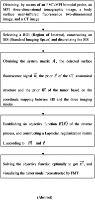
obtaining, by means of an optical/magnetic particle bimodal probe, an Magnetic Particle Imaging (MPI) three-dimensional tomographic image comprising tumor information in a detected living body, a body surface near-infrared fluorescence two-dimensional image, and a Computed Tomography (CT) image comprising anatomical structure information of tissues and organs around a tumor;
taking the tumor, adjacent tissues and organs as a Region of interest (ROI), constructing an Standard Imaging Space (SIS) capable of accommodating the ROI, and discretizing the SIS using a finite element method;
respectively performing threshold segmentation preprocessing on the CT image and the MPI three-dimensional tomographic image to obtain a preprocessed CT image and a preprocessed MPI three-dimensional tomographic image;
encoding the discretized SIS to obtain a position vector x;
mapping the body surface near-infrared fluorescence two-dimensional image to a discretized SIS surface to obtain a detected surface fluorescence signal b;
taking the center coordinate of the discretized SIS as an imaging space center of the CT image, taking each pixel of the preprocessed CT image as a voxel point, obtaining the nearest grid node of the current voxel point in the discretized SIS, giving the properties of the organ corresponding to the current voxel point to the grid node, going through the voxel points corresponding to each pixel, mapping the preprocessed CT image into the discretized SIS, and obtaining the prior c of the anatomical structure of the tissues and organs around the tumor;
arranging registration reference points, adjusting the imaging spatial coordinate system of the MPI three-dimensional tomographic image to be consistent with the imaging spatial coordinate system of the CT image, performing a registration according to the mark points and adjusting the MPI spatial coordinate system to be consistent with the CT imaging space, thereby improving the accuracy and credibility of the registration, adjusting the resolution of the MPI three-dimensional tomographic image and the CT image to the same by adopting an interpolation method or super-resolution method, taking each pixel of the preprocessed MPI three-dimensional tomographic image as a voxel point, obtaining the nearest grid node of the current voxel point in the discretized SIS, giving the magnetic particle concentration at the spatial position corresponding to the current voxel point to the grid node, going through the voxel points corresponding to each pixel, and mapping the preprocessed MPI three-dimensional tomographic image into the discretized SIS, and obtaining the prior m of the tumor;
performing forward model calculation based on SIS after the surface mapping and internal mapping to obtain a linear relationship A between the surface fluorescence signal b and the internal three-dimensional fluorescence distribution;
establishing an objective function E(x) of fluorescence molecular tomography reconstruction based on the linear relationship A and the position vector x;
 wherein λ represents the regularization parameter, L represents the Laplace regularization matrix, ∥•∥22 represents the square of the vector 2 norm, and ∥•∥pp represents the P-power of the vector P norm;
merging the subspaces corresponding to different organs or tissues in the preprocessed CT image and the subspaces corresponding to the positions and shapes of tumors and different organs or tissues in the preprocessed MPI three-dimensional tomographic image to obtain a merged space S;
constructing the Laplace regularization matrix L based on the merged space S,
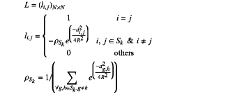 wherein li,j represents the elements in row i and column j of the Laplace matrix, R represents the Gaussian kernel radius, di,j represents the Euclidean distance between the grid node i and the grid node j in the merged space S, dg,h represents the Euclidean distance between the grid node g and the grid node h in the subspace Sk, Sk represents the subspace k in the merged space, N×N represents the dimension of the Laplace matrix, and N represents the number of all discretized points in SIS space;
based on the Laplacian regularization matrix L, selecting an iteration method for solving an objective function, and iteratively solving the objective function E(x) to obtain a fluorescence molecular tomography reconstruction result x*.
|