| CPC A61F 2/966 (2013.01) [A61B 1/005 (2013.01); A61B 1/00082 (2013.01); A61B 1/00087 (2013.01); A61B 1/05 (2013.01); A61B 1/0676 (2013.01); A61B 1/307 (2013.01); A61F 2/9517 (2020.05); A61F 2002/047 (2013.01)] | 15 Claims |
|
1. A method of imaging delivery of an implant, the method comprising:
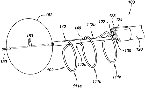
advancing a delivery system within a urethra of a patient, wherein the delivery system comprises an outer tubular member comprising an imaging device located in a distal end region of the outer tubular member, an inner tubular member within the outer tubular member and housing at least a portion of an implant, and one or more structures slidably advanceable within the inner tubular member to cause deployment of the implant from within the inner tubular member, wherein the outer tubular member, inner tubular member, and one or more structures are each coupled with a proximal control device outside of the patient;
longitudinally retracting the inner tubular member with respect to the proximal control device and the one or more structures to at least partially deploy the implant from the inner tubular member; and
while the inner tubular member is being longitudinally retracted, concurrently (a) longitudinally retracting the outer tubular member with respect to the proximal control device and (b) imaging the at least partially deployed implant with the imaging device located at the distal end region of the outer tubular member.
|