| CPC A61B 8/0891 (2013.01) [A61B 5/742 (2013.01); A61B 5/7405 (2013.01); A61B 5/7455 (2013.01); A61B 8/06 (2013.01); A61B 8/085 (2013.01); A61B 8/14 (2013.01); A61B 8/42 (2013.01); A61B 8/4444 (2013.01); A61B 8/4461 (2013.01); A61B 8/463 (2013.01); A61B 8/488 (2013.01); A61B 8/5223 (2013.01); A61B 8/5292 (2013.01); A61B 8/0858 (2013.01)] | 27 Claims |
|
1. An ultrasound system for acquiring a sequence of ultrasound image data sets for the detection of an atheroma in an individual, comprising:
a handheld transducer configured to direct ultrasound signals towards tissue and to detect corresponding echo signals from constituent layers of the tissue;
processing circuitry that is configured to execute program instructions to:
produce ultrasound image data from the detected echo signals and analyze the image data via the processing circuitry to:
(i) dynamically define, without user input, a region of interest from the tissue as the echo signals are being acquired;
(ii) identify an artery from the region of interest as the echo signals are being acquired, based on:
a brightness of a wall of the artery,
a darkness in a lumen of the artery, and
a difference between the brightness of the artery wall and the darkness of the artery lumen; and
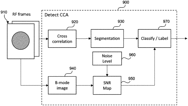
(iii) identify an onset of an atheroma from the identified artery in the region of interest as the echo signals are being acquired by measuring one or more thicknesses of constituent layers of the artery wall,
control a user interface that indicates a direction to the user to manually change the position or orientation of the handheld transducer to position the artery in a desired location in the image data; and
store a sequence of ultrasound image data where the artery is in the desired location in the image data.
|