| CPC A61B 5/361 (2021.01) [A61B 5/316 (2021.01); A61B 5/339 (2021.01); A61B 5/352 (2021.01); A61B 5/7239 (2013.01); A61B 5/7275 (2013.01); A61B 5/743 (2013.01); A61B 5/287 (2021.01); A61B 5/366 (2021.01); A61B 18/1492 (2013.01); A61B 2562/046 (2013.01)] | 19 Claims |
|
1. A method for determining atrial fibrillation regions of interest to be ablated comprising:
acquiring, via a plurality of sensors, electro-cardiogram (ECG) signals over time, each ECG signal acquired via one of the plurality of sensors and representing electrical activity of one of a plurality of different areas of a heart, each ECG signal comprising at least an R wave and an S wave;
determining local activation times (LATs) of each ECG signal, each LAT of a respective ECG signal corresponding to acquired electrical activity at a different point in time;
identifying a plurality of LATs determined to be part of a same cardiac activation cycle;
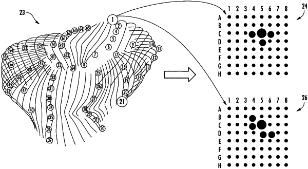
generating first mapping information for visually indicating, at locations on a local activation time (LAT) map for each ECG signal, a number of instances, during a period of time that includes multiple cardiac activation cycles, in which at least one of the plurality of LATs is a chronologically earliest LAT; and
generating second mapping information for visually indicating, at locations on an R-S map corresponding to the same locations on the LAT map, sizes of R-to-S ratios for each ECG signal in the period of time that includes multiple cardiac activation cycles, each R-to-S ratio comprising a ratio of a magnitude of the R wave to a magnitude of the S wave,
wherein, potential focal sources of activation of the heart are identified based on the visually indicated number of times at each location on the LAT map and the visually indicated sizes of the R-to-S ratios at each corresponding location on the R-S map.
|