| CPC G06T 7/40 (2013.01) [G01N 21/25 (2013.01); G06T 3/0068 (2013.01); G06T 7/0012 (2013.01); G06T 7/77 (2017.01); G06T 7/97 (2017.01); A61B 3/12 (2013.01); A61B 5/02007 (2013.01); A61B 5/4088 (2013.01); A61B 5/4381 (2013.01); A61B 5/444 (2013.01); G01N 21/55 (2013.01); G01N 21/6456 (2013.01); G06T 2207/10152 (2013.01); G06T 2207/30024 (2013.01)] | 12 Claims |

|
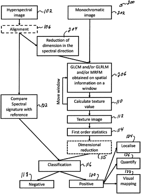
1. A method for imaging a biological tissue, comprising:
obtaining a monochromatic image of the biological tissue;
performing a texture analysis of the biological tissue using spatial information of the monochromatic image of the biological tissue to identity features of the biological tissue;
generating a texture image based on the features of the biological tissue by:
defining a moving window having a k·l size, wherein k is a number of pixels of the moving window defined in a first dimension of the image of the biological tissue and wherein l is a number of pixels of the moving window defined in a second dimension of the image of the biological tissue,
using information from the image of the biological tissue contained in the moving window to calculate a value of a pixel of the texture image,
successively displacing the moving window by a first predetermined number of pixels in the first dimension of the image of the biological tissue,
repeating the calculation of pixel values of the texture image following each displacement of the moving window in the first dimension of the image of the biological tissue,
successively displacing the moving window by a second predetermined number of pixels in the second dimension of the image of the biological tissue, and
repeating the calculations of pixel values of the texture image and the displacements of the moving window over the first dimension of the image of the biological tissue following each displacement of the moving window over the second dimension of the image of the biological tissue; and
classifying the biological tissue of the subject as normal or abnormal at least in part based on a comparison between first order statistics of the texture image and predetermined values.
|