| CPC A61B 5/0073 (2013.01) [A61B 5/0075 (2013.01); G06N 3/084 (2013.01); G06N 5/046 (2013.01); G06N 20/00 (2019.01); G06T 11/006 (2013.01); G06T 2210/41 (2013.01)] | 2 Claims |

|
1. A near-infrared spectroscopy tomography reconstruction method based on neural network applied to biological tissue, comprising: an energy diffusion approximation equation (1) of light in the biological tissue is expressed as:
 wherein, c represents a speed at which light travels through the tissue, t represents time, r represents a coordinate position vector, κ represents scattering coefficient, μa represents absorption coefficient; Φ(r, t) represents a photon fluence rate at position r; q0(r,t) represents a light source; an energy diffusion approximation equation (2) under continuous wave mode is adopted without considering an influence of time on the energy diffusion approximation equation (1):
−∇·κ(r)∇Φ(r)+μa(r)Φ(r)=q0(r) (2)
wherein q0(r) represents an isotropic light source; Φ(r) represents photon density distribution at position r;
a boundary condition corresponding to steady-state diffusion equation in near-infrared optical tomography is: air tissue boundary is expressed by exponential mismatch type III condition which is also known as Robin or mixed boundary condition which is expressed as:
Φ(ξ)+2An(ξ)n·κ(ξ)∇Φ(ξ)=0 (3)
wherein ξ represents point on an outer boundary of tissue; n represents a unit outer normal; An depends on mismatched relative refractive index (RI) between tissue and air and is expressed as:
 wherein Rn represents internal reflection coefficient of diffusion transmission; n is related to optical refraction coefficient deviation inside and outside the outer boundary;
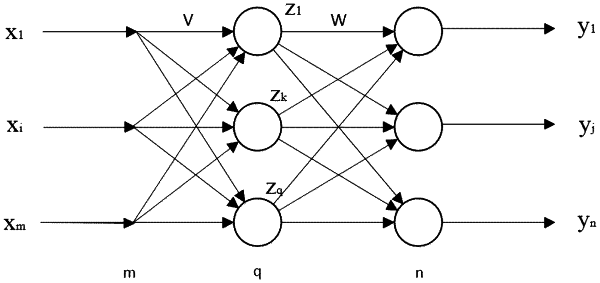
based on a distribution of optical parameters, finite element method (FEM) and light approximate transmission equation is adopted to obtain outer boundary measured value Φ, Φ is used as an input of back propagation (BP) neural network x, then an output y of the BP neural network is a corresponding optical parameter distribution;
the near-infrared spectroscopy tomography reconstruction method further comprises:
establishing circular replica and finishing finite element mesh generation through Matlab toolbox nirfast; wherein a plurality of light sources and light detectors are set up along an outer boundary of the circular replica, measurements are obtained through the light sources and the light detectors, and imaging pixels are uniform finite element nodes on the circular replica; measured value and absorption coefficient of each finite element node are taken as the input and the output of the BP neural network, respectively;
data set used for training of the BP neural network is calculated through forward solving process of nirfast toolbox's; there are a plurality of samples in the data set, including samples in a training set and samples in a test set, each sample contains an abnormal region, the abnormal region is a circular region with a radius, and a center position of the abnormal region for different sample is different.
|