| CPC A61B 8/0866 (2013.01) [A61B 8/0883 (2013.01); A61B 8/463 (2013.01); A61B 8/5223 (2013.01); G06T 7/0012 (2013.01); G06T 11/001 (2013.01); G06T 2200/24 (2013.01); G06T 2207/10016 (2013.01); G06T 2207/10132 (2013.01); G06T 2207/20036 (2013.01); G06T 2207/20084 (2013.01); G06T 2207/30044 (2013.01); G06T 2207/30048 (2013.01); G06T 2207/30101 (2013.01)] | 30 Claims |

|
1. A computer implemented method for analysis of fetal ultrasound images, the method comprising:
receiving a plurality of sets of image data generated by an ultrasound system during a fetal ultrasound examination, each set of image data of the plurality of sets of image data comprising a plurality of frames;
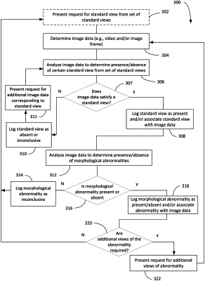
analyzing a set of image data of the plurality of sets of image data to automatically determine that one or more frames of the set of image data corresponds to a standard view of a plurality of standard views;
analyzing the set of image data to automatically determine that the one or more frames is indicative of a first morphological abnormality of a plurality of morphological abnormalities;
generating a user interface for display, wherein the user interface comprises:
(a) an image data viewer adapted to visually present the set of image data;
(b) a standard view indicator corresponding to the set of image data presented on the image data viewer and visually indicating whether each standard view of the plurality of standard views is present in the set of image data; and
(c) a morphological anomaly indicator corresponding to the set of image data presented on the image data viewer and visually indicating whether each morphological abnormality of the plurality of morphological abnormalities is present in the set of image data,
wherein the standard view indicator indicates that a first standard view is present in the set of image data and the morphological anomaly indicator indicates a first morphological abnormality is present when the image data viewer visually presents the set of image data.
|