| CPC A61B 8/5246 (2013.01) [A61B 8/08 (2013.01); A61B 8/461 (2013.01); A61B 8/483 (2013.01)] | 11 Claims |
|
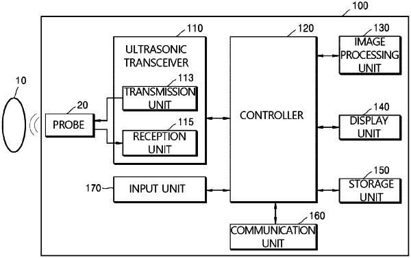
1. A method of displaying an ultrasonic image, the method comprising:
acquiring, by an ultrasonic transceiver, first data by transmitting a first ultrasonic pulse to an object and by receiving an echo signal reflected from the object;
acquiring, by the ultrasonic transceiver, a plurality of two-dimensional data sets by repeatedly performing, a plurality of times at predetermined time intervals, an operation of acquiring a two-dimensional data set representing a cross-section of the object by transmitting second ultrasonic pulses different from the first ultrasonic pulse to the object and receiving echo signals of the second ultrasonic pulses reflected from the object, wherein each of the second ultrasonic pulses includes an asymmetric pulse in which a negative pressure component is dominant as compared to a positive pressure component;
determining, by a controller, from among regions of the cross-section of the object represented by the plurality of two-dimensional data sets, a region on which a variance of a phase change over time of the echo signals of the second ultrasonic pulses acquired at the plurality of times in the plurality of two-dimensional data sets is larger than or equal to a predetermined value as a microcalcified tissue region; and
displaying, by a display unit, an ultrasonic image representing the cross-section of the object based on the first data and an image indicating the determined microcalcified tissue region.
|