| CPC A61B 1/043 (2013.01) [A61B 1/00009 (2013.01); A61B 1/00045 (2013.01); A61B 1/00082 (2013.01); A61B 1/00186 (2013.01); A61B 1/05 (2013.01); A61B 1/0638 (2013.01); A61B 1/0676 (2013.01); A61B 1/0684 (2013.01); A61B 5/004 (2013.01); A61B 5/0044 (2013.01); A61B 5/0084 (2013.01); A61B 5/14503 (2013.01); A61B 5/14546 (2013.01); A61B 5/1459 (2013.01); A61B 5/6853 (2013.01); A61B 18/00 (2013.01); A61B 18/1492 (2013.01); A61B 90/30 (2016.02); A61B 90/35 (2016.02); A61B 90/361 (2016.02); A61B 2018/0022 (2013.01); A61B 2018/0212 (2013.01); A61B 2034/301 (2016.02); A61B 2090/365 (2016.02); Y02A 90/10 (2018.01)] | 20 Claims |

|
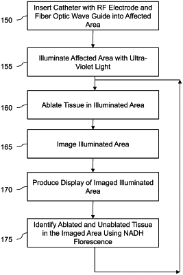
1. A system for imaging tissue, the system being configured for use in connection with tissue ablation, comprising:
a light source providing light for illuminating a tissue to excite mitochondrial nicotinamide adenine dinucleotide hydrogen (NADH) in the tissue;
a sensor for detecting NADH fluorescence from the illuminated tissue; and
a processor programmed to perform the steps of:
obtaining the detected NADH fluorescence from the sensor during ablation of the tissue,
generating a digital representation of the detected NADH fluorescence for monitoring a progression of the ablation of the tissue, and
while the tissue is being ablated, determining a decrease in the detected NADH fluorescence and updating the digital representation to show the measured decrease in the detected NADH fluorescence that is indicative of the progression of the ablation of the tissue.
|