| CPC G06T 7/0012 (2013.01) [A61B 1/000094 (2022.02); G06T 7/11 (2017.01); G06T 7/136 (2017.01); G06T 7/168 (2017.01); G06T 2207/20024 (2013.01); G06T 2207/20081 (2013.01); G06T 2207/20084 (2013.01); G06T 2207/20132 (2013.01); G06T 2207/30096 (2013.01)] | 30 Claims |

|
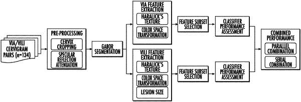
1. A method for automated detection of cervical pre-cancer, the method comprising:
providing at least one cervigram;
pre-processing the at least one cervigram including automatically segmenting a region from the cervix for further analysis;
extracting texture-based features from the segmented region of the at least one pre-processed cervigram; and
classifying the at least one cervigram as negative or positive for cervical pre-cancer based on the extracted features.
|
|
13. A method for developing an algorithm for automated cervical cancer diagnosis, the method comprising:
providing a plurality of cervigrams;
pre-processing each cervigram including automatically segmenting a region from the cervix for further analysis;
extracting texture-based features from the segmented region of each pre-processed cervigram; and
establishing a classification model based on the extracted features for each cervigram,
wherein the classification model is configured to classify additional cervigrams as negative or positive for cervical pre-cancer.
|