| CPC A61B 5/14552 (2013.01) [A61B 5/4848 (2013.01); A61B 5/7221 (2013.01); A61B 5/7275 (2013.01); A61B 5/742 (2013.01); A61B 5/743 (2013.01); A61B 5/7475 (2013.01); A61B 2560/0214 (2013.01); A61B 2560/0425 (2013.01); A61B 2560/0475 (2013.01); A61B 2562/0238 (2013.01); A61B 2562/0242 (2013.01); A61B 2562/046 (2013.01)] | 21 Claims |

|
1. A method comprising:
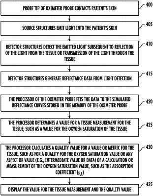
providing an oximeter device, wherein the oximeter device is wireless;
providing data points for simulated reflectance curves that are accessible to the oximeter device;
using the oximeter device, making an oxygen saturation measurement of a tissue comprising emitting light from a source of the oximeter device into the tissue, and receiving light reflected from the tissue at detectors of the oximeter device in response to the emitted light, wherein a plurality of detector responses is obtained for the reflected light;
fitting the detector responses to the data points for the simulated reflectance curves to determine an absorption coefficient value for the tissue;
using the absorption coefficient value, determining an oxygen saturation value for the tissue;
calculating an error value from the fitting of the detector responses to the simulated reflectance curves, and using the error value, determining a quality metric value for the oxygen saturation value; and
displaying on a screen the oxygen saturation value and the quality metric value.
|