| CPC A61B 3/1233 (2013.01) [A61B 3/0025 (2013.01); A61B 3/102 (2013.01); A61B 3/1241 (2013.01); A61B 3/165 (2013.01); G06T 7/0016 (2013.01); A61B 8/10 (2013.01); G06T 2207/10101 (2013.01); G06T 2207/30041 (2013.01); G06T 2207/30104 (2013.01)] | 18 Claims |

|
1. A method, comprising:
capturing, using an optical coherence tomography instrument, imaging data of a retina of an eye, wherein the imaging data includes first topographical data and second topographical data, wherein the second topographical data is captured subsequent the first topographical data, wherein the imaging data is associated with an imaged volume of the eye, wherein the first topographical data is captured at a first intraocular pressure and the second topographical data is captured at a second intraocular pressure or the imaging data is captured at the first intraocular pressure and additional imaging data associated with the imaged volume of the eye is captured at a second intraocular pressure, wherein the first intraocular pressure is different from the second intraocular pressure, and wherein the imaging data further includes first intraocular pressure data related to the first intraocular pressure and second intraocular pressure data related to the second intraocular pressure or the imaging data further includes first intraocular pressure data related to the first intraocular pressure and the additional imaging data includes second intraocular pressure data related to the second intraocular pressure;
identifying, using a processing system of or in data communication with the optical coherence tomography instrument, vasculature using the imaging data, wherein identifying vasculature includes comparing the first topographical data to the second topographical data to identify voxels in the imaging data that change over time;
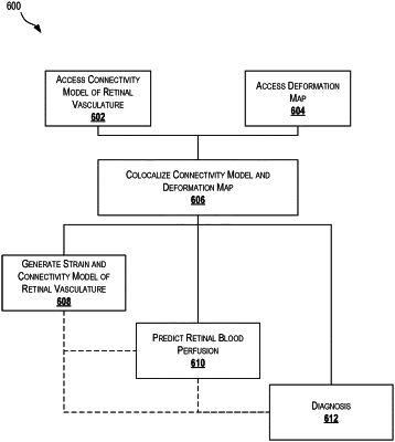
generating a connectivity model of vasculature of the retina using the identified vasculature, wherein the connectivity model of vasculature corresponds to a three-dimensional graph representative of a vessel tree of the vasculature;
calculating deformation data for at least a portion of the imaged volume of the eye based on the first intraocular pressure data and the second intraocular pressure data;
generating a deformation map using the deformation data, wherein the deformation map is a data structure corresponding to a three-dimensional map of deformation intensity, and wherein the deformation map describes amounts of strain occurring at one or more voxels between the first intraocular pressure and the second intraocular pressure;
colocalizing the connectivity model and the deformation map to generate a colocalized connectivity model and deformation map identifying regions of the connectivity model that undergo various amounts of strain during changes in the intraocular pressure; and
predicting retinal blood perfusion using the colocalized connectivity model and deformation map, wherein the predicting comprises supplying different conditions including changes in intraocular pressure and changes in systemic pressure to the colocalized connectivity model and deformation map to predict how the vasculature would react to the different conditions and estimate a quantification of the retinal blood perfusion given the different conditions.
|