| CPC G06T 7/11 (2017.01) [G06T 7/0012 (2013.01); G16H 30/40 (2018.01); G16H 50/20 (2018.01); G06T 2207/10116 (2013.01); G06T 2207/20081 (2013.01); G06T 2207/20084 (2013.01); G06T 2207/30008 (2013.01); G06T 2207/30061 (2013.01); G06T 2207/30168 (2013.01)] | 18 Claims |

|
1. A method for processing medical chest images, the method comprising:
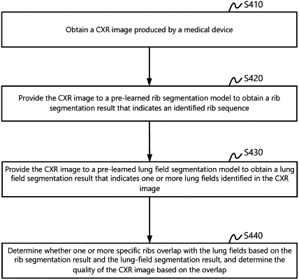
obtaining a chest image;
segmenting the chest image based on a machine-learned rib segmentation model to obtain a rib segmentation result, wherein the rib segmentation result indicates a rib sequence identified in the chest image, and wherein segmenting the chest image based on the machine-learned rib segmentation model comprises:
segmenting the chest image to determine a first region that is estimated to enclose a plurality of ribs identified in the chest image;
segmenting the first region to determine one or more second regions, wherein each of the one or more second regions is associated with a respective subset of the plurality of ribs enclosed in the first region; and
segmenting each of the one or more second regions to identify individual ribs located in the each of the one or more second regions;
segmenting the chest image based on a machine-learned lung field segmentation model to obtain a lung field segmentation result, wherein the lung field segmentation result indicates one or more lung fields identified in the chest image;
determining, whether a predetermined set of one or more specific ribs overlaps with the one or more lung fields according to the rib segmentation result and the lung field segmentation result; and
determining a quality of the chest image in accordance with whether the predetermined set of one or more specific ribs overlaps with the one or more lung fields.
|