| CPC A61B 34/20 (2016.02) [A61B 8/0841 (2013.01); A61B 8/4488 (2013.01); A61B 8/463 (2013.01); A61B 8/485 (2013.01); A61B 8/5215 (2013.01); A61B 10/0275 (2013.01); A61B 17/3403 (2013.01); A61B 34/25 (2016.02); A61B 90/39 (2016.02); G01S 7/52073 (2013.01); G01S 15/8906 (2013.01); G06N 20/00 (2019.01); G16H 10/40 (2018.01); G16H 20/40 (2018.01); G16H 30/40 (2018.01); G16H 40/63 (2018.01); A61B 8/0825 (2013.01); A61B 8/481 (2013.01); A61B 2017/3413 (2013.01); A61B 2034/2063 (2016.02); A61B 2034/2065 (2016.02); A61B 2090/378 (2016.02); A61B 2090/3925 (2016.02); A61B 2090/3983 (2016.02)] | 15 Claims |
|
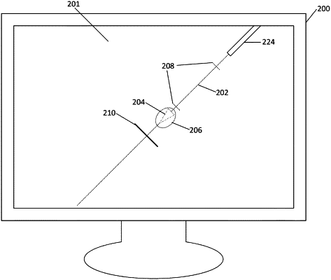
1. A method for providing guidance for operation of a biopsy needle, the method comprising:
emitting an array of ultrasonic sound waves from an ultrasonic transducer of an ultrasound probe;
detecting reflected ultrasonic sound waves by the ultrasonic transducer, wherein the reflected ultrasonic sound waves include at least a portion of the array of ultrasonic sound waves after being reflected from an interior of a patient;
generating image data from the reflected ultrasonic sound waves;
identifying, by a processor, within the generated image data, at least a portion of the biopsy needle within the interior of the patient;
based at least in part on the identification the portion of the biopsy needle, determining, by the processor, a predicted location of an aspect of the biopsy needle based at least in part on one or more biopsy needle properties stored in memory operatively connected to the processor, the biopsy needle properties comprising at least one of a needle length, a needle gauge, a needle wall thickness, a needle material composition, a needle tip geometry, and a needle firing mechanism; and
displaying, on a display operatively connected to the processor, an ultrasound image based on the generated image data;
determining a deflection probability for a post-fire needle tip location based on at least one of: (1) experimental data for the type of biopsy needle and (2) the one or more stored properties of the biopsy needle; and
displaying a deflection probability indicator on the ultrasound image, wherein the deflection probability indicator indicates a range for the post-fire needle tip locations based on the determined deflection probability, wherein the deflection probability indicator depicts a probability distribution for the post-fire needle tip location;
wherein the deflection probability indicator is based at least in part on a standard deviation of the post-fire needle tip locations.
|