| CPC A61B 3/12 (2013.01) [A61B 3/0008 (2013.01); A61B 3/0033 (2013.01); A61B 3/158 (2013.01)] | 21 Claims |

|
1. An ophthalmic diagnostic system for imaging an eye, comprising:
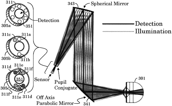
a detector configured for capturing an image of the eye, the detector having a detector aperture;
at least a first light source and a second light source proximate to the detector aperture; and
one or more data processors;
wherein:
the detector configured to capture a first image of the eye with the first light source actuated and the second light source not actuated, and configured to capture a second image of the eye with the second light source actuated and the first light source not actuated and configured to generate output signals in response thereto; and
the one or more data processors are configured to use the output signals to extract a first section of the first image excluding reflex artifacts from the first light source, extract a second section of the second image excluding reflex artifacts from the second light source, and combine the first and second sections to construct a composite image.
|