| CPC G01R 33/481 (2013.01) [A61B 6/037 (2013.01); A61B 6/0407 (2013.01); A61B 6/4417 (2013.01); G01R 33/4812 (2013.01); G01T 1/2985 (2013.01); G06T 11/003 (2013.01); A61B 6/481 (2013.01); A61B 6/5235 (2013.01); G06T 2207/10088 (2013.01); G06T 2207/10104 (2013.01)] | 17 Claims |

|
1. A method for generating transmission information in a time-of-flight positron emission tomography (PET) scanner having a patient tunnel and a plurality of PET detector rings wherein the PET scanner uses continuous bed motion to move a patient bed having a patient through the patient tunnel and wherein the patient receives a positron-emitting radioisotope dose prior to undergoing a PET scan, comprising:
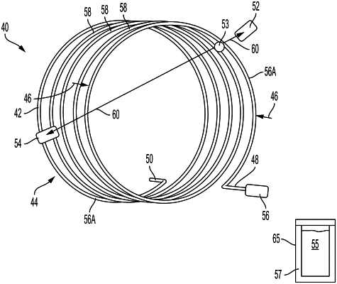
storing the positron-emitting radioisotope in a radiation shielded container element;
moving the radioisotope into a stationary vessel located adjacent to the PET detector rings and within a field of view of the PET scanner at substantially the same time that the patient receives the radioisotope dose to form a stationary transmission source wherein the transmission information is generated while the patient bed undergoes the continuous bed motion; and
withdrawing the radioisotope from the vessel when the PET scan is complete and storing the radioisotope in the container element wherein the PET scanner is integrated with an x-ray computed tomography (CT) scanner that generates a CT based attenuation map that is algorithmically compared to a PET based attenuation map wherein the PET based attenuation map is used to replace the CT based attenuation map in PET image reconstruction if a discrepancy occurs between the CT and PET based attenuation maps.
|