| CPC G06T 7/0016 (2013.01) [G06N 3/08 (2013.01); G06T 7/38 (2017.01); G16H 30/20 (2018.01); G06T 2207/10088 (2013.01); G06T 2207/20081 (2013.01); G06T 2207/20084 (2013.01); G06T 2207/20224 (2013.01)] | 21 Claims |
|
1. A computer-implemented method for identifying pathological changes in follow-up medical images, the computer-implemented method comprising:
providing reference image data showing a body part of a patient at a first time;
providing follow-up image data showing the body part of the patient at a second time, the second time being after the first time;
generating one or more first deformation fields for the reference image data and the follow-up image data using at least one image registration, the one or more first deformation fields describing anatomical deformations in the body part between the reference image data and the follow-up image data;
aligning the reference image data and the follow-up image data using the one or more first deformation fields to generate co-aligned image data; and
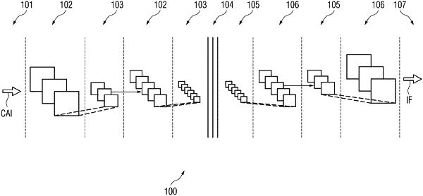
analyzing the co-aligned image data to identify pathological changes using a machine learned network, the pathological changes occurring in the body part of the patient between the first time and the second time, and the machine learned network being trained to identify pathological changes in the co-aligned image data.
|