| CPC A61B 8/145 (2013.01) [A61B 8/4281 (2013.01); A61B 8/4455 (2013.01); A61B 8/4494 (2013.01); A61B 8/5215 (2013.01)] | 8 Claims |
|
1. A spiral ultrasonic tomography imaging method with an ultrasonic probe, the ultrasonic probe comprises a plurality of array elements, the spiral ultrasonic tomography imaging method, comprising:
under a premise of a uniform displacement of the ultrasonic probe along a Z axis, using synchronous rotation of the array elements or performing cyclic switching of a change trajectory linked by positions of an emission array element selected from one of the array elements in the ultrasonic probe according to a preset fixed spiral direction or a preset changing spiral direction each time, so that the change trajectory of positions of the emission array element over time in a three-dimensional space at each ultrasonic emission time is distributed along a spiral line or along a curve composed of a plurality of parts of the spiral line; and
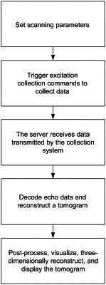
during a process of the uniform displacement of the ultrasonic probe along the Z axis, emitting, by the ultrasonic probe, an ultrasonic wave and receiving and collecting, by the ultrasonic probe, echo data, and the echo data been collected is stored and post-processed to realize ultrasonic tomography imaging of an object to be detected, wherein
the array elements of the ultrasonic probe are located outside the object to be detected, each time the ultrasonic wave is emitted, one of the array elements serves as the emission array element, and the other array elements all serve as receiving array elements, each array element of the ultrasonic probe has functions of the emission array element and a receiving array element among the receiving array elements, and the echo data received by each of the receiving array elements carries tomography imaging information of a local area of the object to be detected near a plane where the ultrasonic probe is located;
wherein the ultrasonic probe is a concave-array ultrasonic probe with the plurality of array elements, the ultrasonic probe performs at least one of steps (a) and (b) or a combination thereof:
(a) performing, by the ultrasonic probe, the uniform displacement along the Z axis along a preset Z-axis direction and at a preset displacement rate while the emission array element in the ultrasonic probe is maintained to be cyclically switched according to the preset fixed spiral direction, and a frequency of switching the emission array element among the array elements satisfies a preset requirement;
(b) performing, by the ultrasonic probe, the uniform displacement along the Z axis along the preset Z-axis direction and at the preset displacement rate while the emission array element in the ultrasonic probe is maintained to be cyclically switched according to the preset changing spiral direction, the frequency of switching the emission array element among the array elements satisfies the preset requirement, and a frequency of changes in the preset fixed spiral direction or in the preset changing spiral direction also satisfies the preset requirement, and
wherein performing data post-processing by:
rearranging and encoding the echo data been stored, so that the echo data is arranged according to an order received by the ultrasonic probe;
computing and reconstructing to obtain each tomogram;
sequentially arranging each tomogram to perform three-dimensional reconstruction, and;
performing logarithmic compression and color or gray-scale mapping to obtain a three-dimensional image, wherein the tomogram is one of a reflection reconstruction tomogram, a sound velocity reconstruction tomogram, and an attenuation coefficient reconstruction tomogram, or a combination thereof.
|