| CPC A61B 8/06 (2013.01) [A61B 8/461 (2013.01); A61B 8/5246 (2013.01); G01S 15/8906 (2013.01); G01S 15/8979 (2013.01); A61B 6/547 (2013.01); A61B 8/5207 (2013.01); G01S 15/8927 (2013.01)] | 18 Claims |

|
1. An ultrasound grayscale flow imaging system, comprising:
a probe;
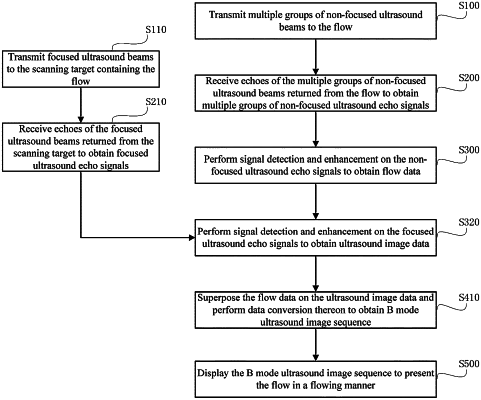
a transmitting circuit which excites the probe to transmit multiple groups of non-focused ultrasound beams and focused ultrasound beams to a scanning target containing a flow, wherein the non-focused ultrasound beams and focused ultrasound beams comprise only B-mode pulses;
a receiving circuit which receives echoes of the multiple non-focused ultrasound beams returned from the flow to obtain multiple groups of non-focused ultrasound echo signals and receives echoes of the multiple focused ultrasound beams returned from the scanning target to obtain multiple groups of focused ultrasound echo signals;
a signal processing unit which obtains grayscale flow data according to the multiple groups of non-focused ultrasound echo signals using grayscale flow imaging without using Doppler processing;
a B-mode signal processing unit which obtains grayscale ultrasound image data according to the multiple groups of focused ultrasound echo signals;
an image processing unit which overlays the grayscale flow data on the grayscale ultrasound image data and obtains a B-mode ultrasound image sequence according to the grayscale ultrasound image data overlaid with the grayscale flow data, wherein the image processing unit is further configured to:
calculate variances of I data and Q data demodulated from the grayscale flow data, wherein the variances are calculated according to the formula,
 where i represents a sampling time, Ii represents I data at ith time, and Qi represents Q data at ith time;
determine non-flow regions and flow regions based on a variance threshold; and
map trends of the variances with grayscales and/or colors to obtain the B-mode ultrasound image sequence overlaid with the grayscales and/or colors or obtain the B-mode ultrasound image sequence overlaid with the grayscales and/or colors based on regions; and
a display device which displays the B-mode ultrasound image sequence.
|