| CPC A61B 3/152 (2013.01) [A61B 3/0008 (2013.01); H04N 23/58 (2023.01)] | 9 Claims |

|
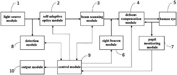
1. A large field-of-view adaptive optics retinal imaging system with common optical path beam scanning, characterized in that, the system comprises: a light source module, an adaptive optics module, a beam scanning module, a defocus compensation module, a sight beacon module, a pupil monitoring module, a detection module, a control module and an output module;
the light source module is configured to emit a parallel light beam, wherein the parallel light beam irradiates a human eye after sequentially going through the adaptive optics module, the beam scanning module and the defocus compensation module, imaging light that is scattered by the human eye and carries aberration information of the human eye and light intensity information returns along an original optical path of the parallel light beam and reaches the adaptive optics module and the detection module;
the adaptive optics module is configured to receive the imaging light carrying the aberration information of the human eye, and perform real-time measurement and correction of aberration of the human eye;
the beam scanning module is controlled by the control module, and the beam scanning module is configured in different scanning modes for carrying out different scanning imaging functions at least including a large field-of-view imaging function, a small field-of-view high-resolution imaging function and a large field-of-view high-resolution imaging function;
the defocus compensation module is configured to achieve compensation of refractive error of the human eye;
the sight beacon module is configured to guide and fix a sight beacon in different areas of a retina of the human eye;
the pupil monitoring module is configured to align and monitor a pupil of the human eye;
the detection module is configured to acquire the returning imaging light of the human eye, convert the imaging light into an electrical signal, and transmit the electrical signal to the control module; and
the output module is configured to connect to the control module, and display and store imaging images of the human eye;
wherein the beam scanning module is configured to comprise a first scanning mirror and a second scanning mirror, and the two scanning mirrors are connected through a transmissive or reflective telescope to achieve pupil phane matching;
the first scanning mirror is configured to perform transverse scanning of the retinal plane, and the second scanning mirror is configured to perform vertical scanning of the retinal plane under the driving of a periodic voltage, in order to carry out the large field-of-view imaging function;
the second scanning mirror is configured to generate a certain transverse and vertical inclination angle under the driving of a DC voltage, the second scanning mirror is also configured to perform transverse and vertical two-dimensional scanning of the retinal plane under the driving of a periodic voltage at the same time of generating the transverse and vertical inclination angle under the driving of a DC voltage, in order to carry out the small field-of-view high-resolution imaging function and the large field-of-view high-resolution imaging function;
wherein the control miodule is configured to control the first scanning mirror and the second scanning miorror in the beam scanning module by outputting a voltage signal to carry out different scanning imaging functions;
wherein, the large field-of-view imaging function is performed by the following process:
the adaptive optics module is in a shutdown state or a non-working power-on state;
the first scanning mirror performs the transverse scanning of the retinal plane under the driving of a periodic voltage signal; the second scanning morror performs the vertical scanning of the retinal plane under the driving of a periodic voltage signal; the retinal scanning angles of the first scanning mirror and the second scanning mirror driven by periodic voltage signals are no less than 20 degrees;
the detection module converts the acquired light signal of the fundus retinal into an electrical signal, the control module synchronized the periodic driving voltage signals of the first scanning mirror and the second scanning mirror, and the control module samples the electrical signal to reconstruct an imaging image of the retina with a large field of view which is then output to the output module for display and storage;
wherein, the small field-of-view high-resolution imaging function is performed by the following process:
the adaptive optics module is in a power-on working state to measure and correct wavefront aberration;
the first scanning mirror performs the transverse scanning of the retinal plane under the driving of a periodic voltage signal; the second mirror generates a certain transverse and vertical inclination angle under the driving of a DC voltage signal for locating the light beam illuminating the fundus retina at a positioin of interest, and then is driven by a periodic voltage signal to perform the vertical scanning of the retinal plane; the retinal scanning angles of the first scanning mirror and the second scanning mirror driven by the periodic voltage signals are no greater than 5 degrees;
the DC voltage signal is calculated by the control module according to a fundus retinal coordinate position;
the detection module converts the acquired light signal of the fundus retina into an electrical signal, the control module synchronizes the periodic driving voltage signals of the first scanning mirror and the second scanning mirror, and the control module samples the electrical signal to reconstruct an imaging image of the retina with a small field of view and high resolution and at the same time marks the fundus retinal coordinate position in the imaging image; the imaging image of the retina with a small field of view and high resolution is output by the control module to the output module for display and storage;
wherein, the large field-of-ivew high-resolution imaging function is performed by the following process:
the adaptive optics module is in a power-on working state to measure and correct wavefront aberration;
the first scanning mirror performs the transverse scanning of the retinal plane under the driving of a periodic voltage signal; the second scanning mirror performs the vertical scanning of the retinal plane under the driving of a periodic voltage signal; the retinal scanning angles of the first scanning mirror and the second scanning mirror driven by periodic voltage signals are no greater that 5 degrees;
at this time, the second scanning mirror generates a certain transverse and vertical inclination angle under the driving of a DC voltage signal to tilt the light beam to sequentially illuminate each area of the fundus retina; a single-time transverse and vertical inclination angle of the second scanning mirror is no greater than 3 degrees, a maximum retinal transverse and longitudinal inclination angle of the second scanning mirror driven by a DC voltage signal is no greater that 15 degrees, the DC voltage signal is calculated by the control module according to a fundus retinal coordinate postion;
when each area of the fundus retina is sequentially illuminated by the light beam, the control module can obtain high-resolution imaging images of each area of the retina, and the control module stitches the high-resolution imaging images according to the fundus retinal coordinate positions of the high-resolution imaging images of the respective areas to obtain an image of the fundus retina with a large field of view and high resolution which is then output to the output module for display and storage.
|