| CPC G16H 30/40 (2018.01) [G06V 10/255 (2022.01); G16H 70/60 (2018.01); H04W 4/80 (2018.02); G06F 2218/10 (2023.01)] | 19 Claims |
|
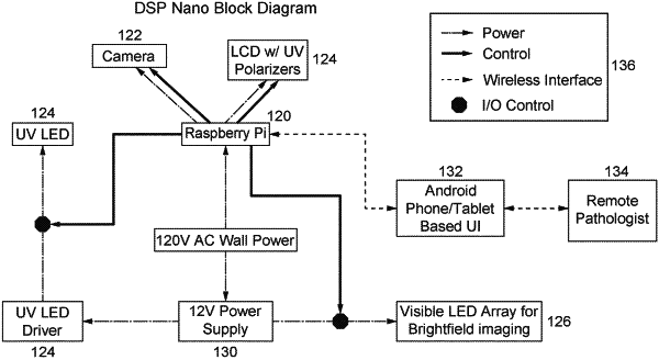
1. A digital spatial profiling (DSP) system comprising:
a housing or other structure for containing at least one component of the DSP system, comprising:
a power source;
an ultraviolet (UV) light source (UVS);
a visible light source (VLS) for bright field imaging;
a chamber comprising a slot configured to receive at least a portion of a slide having a tissue sample thereon;
a personal mobile computing device (PMD) comprising a user interface and a camera sensor, the PMD being arranged relative to the chamber in order to image the tissue sample on the slide within the chamber;
photomasking means configured to selectively illuminate the tissue sample with UV light from the UVS and/or visible light from the VLS;
optic means configured to at least one of direct and/or focus the UVS and/or VLS toward at least one of the tissue sample, the chamber, the photomasking means, and the camera sensor; and
processing circuitry configured to
acquire, via the camera sensor, an image of the tissue sample,
display, via the user interface of the PMD, the image of the tissue sample,
receive, via the user interface of the PMD, user input regarding a region of interest (ROD of the tissue sample, and
illuminate, using the photomasking means, the ROI with UV light,
wherein the PMD is removably attachable to the housing and/or the chamber.
|