| CPC G06T 7/0016 (2013.01) [A61B 90/20 (2016.02); A61B 90/361 (2016.02); G06T 2207/10056 (2013.01); G06T 2207/30024 (2013.01); G06T 2207/30044 (2013.01)] | 11 Claims |

|
1. A method, comprising:
capturing an image of a fertilized egg being cultured such that images of the fertilized egg are captured with a predetermined interval; and
causing a computer to perform a method for analyzing an image of the fertilized egg, comprising:
detecting a male pronucleus and a female pronucleus in the images of the fertilized egg captured;
measuring areas of the female pronucleus and the male pronucleus comprising measuring a diameter in a horizontal direction and a diameter in a vertical direction of the female pronucleus and the male pronucleus, and
calculating respective areas of the male pronucleus and the female pronucleus according to the following formula:
area of pronucleus=π×½×diameter in horizontal direction×½×diameter in vertical direction (μm2), by designating the maximum width in the horizontal direction of the pronucleus as the diameter in the horizontal direction, and designating the maximum width in the vertical direction as the diameter in the vertical direction,
wherein the areas of the female pronucleus and the male pronucleus are measured over time with intervals of every 5 minutes to every 1 hour,
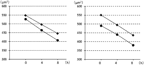
measuring a first area difference between the female pronucleus and the male pronucleus from an image of a fertilized egg captured at a point of time in a period from 1 hour before to 10 hours before a time of occurrence of male and female pronuclear membrane breakdown;
measuring a second area difference between the female pronucleus and the male pronucleus from an image of a fertilized egg captured at a point of time in a period from immediately before to 20 minutes before the time of occurrence of the male and female pronuclear membrane breakdown;
selecting a fertilized egg to be transplanted into the uterine cavity based on measured values of the first area difference and the second area difference, wherein the selecting comprises selecting a fertilized egg that satisfies that the second area difference is less than 40 μm2, and
transplanting the fertilized egg into the uterine cavity.
|