| CPC G06T 7/0012 (2013.01) [A61B 5/4836 (2013.01); G06T 7/11 (2017.01); G06T 7/149 (2017.01); G06T 2207/10088 (2013.01); G06T 2207/20084 (2013.01); G06T 2207/30016 (2013.01); G06T 2207/30096 (2013.01); G06T 2207/30104 (2013.01)] | 28 Claims |

|
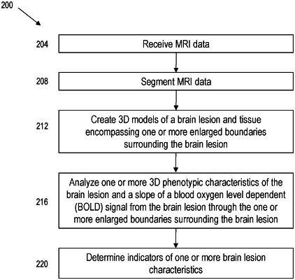
1. A system for determining characteristics of a brain lesion and tissue encompassing boundaries surrounding the brain lesion in three dimensions, the system comprising:
a computer system including at least one processor configured to:
receive data from a magnetic resonance imaging (MRI) machine configured to generate one or more series of images corresponding to a structural and a functional characteristic of a brain lesion and tissue encompassing one or more enlarged boundaries surrounding the brain lesion, the brain lesion having an outer boundary and at least part of the one or more enlarged boundaries surrounding the brain lesion being offset by a given distance from the outer boundary of the brain lesion;
segment the received data to isolate a portion of the received data corresponding to the brain lesion and the tissue surrounding the brain lesion within the one or more enlarged boundaries;
create, based on the segmented data, one or more three-dimensional (3D) models of the brain lesion and the tissue surrounding the brain lesion within the one or more enlarged boundaries;
analyze, based on the one or more 3D models, one or more 3D phenotypic characteristics of the brain lesion and a slope of a blood oxygen level dependent (BOLD) signal from within the brain lesion through the one or more enlarged boundaries, and
determine, based on the one or more 3D phenotypic characteristics and the slope, indicators of one or more characteristics selected from a group of characteristics consisting of: lesion age, extent of injury, remyelination capacity, tissue integrity within the brain lesion, tissue integrity within tissue surrounding the brain lesion, and metabolic activity of the brain lesion within tissue surrounding the brain lesion.
|