| CPC A61B 5/489 (2013.01) [A61B 5/004 (2013.01); G06V 10/56 (2022.01); G06V 10/751 (2022.01); G06V 40/10 (2022.01); H04N 23/55 (2023.01); H04N 25/534 (2023.01); G06V 40/14 (2022.01)] | 8 Claims |

|
1. A camera having blood optical imaging function, comprising:
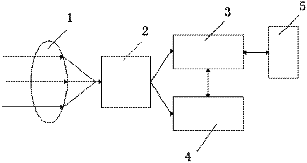
a lens (1), an image sensor (2), a main control module (3), a blood optical imaging algorithm module (4) and an IP network interface module (5);
the lens (1) is located at an image acquiring position of the camera;
an output end of the image sensor (2) are in electric connection with input ends of the main control module (3) and the blood optical imaging algorithm module (4), respectively;
the main control module (3) is in electric connection with the blood optical imaging algorithm module (4) and the IP network interface module (5), respectively;
the blood optical imaging algorithm module (4) is used for extracting continuously changed dynamic optical images of the face blood vessels from the RGB digital image signals, and send the same into the main control module (3) for packaging into IP data packets, ensuring a full extraction of the images; and
the calculation method of the blood optical imaging algorithm module (4) comprises the following steps:
S01: continuously reading the data of N frames of RGB image from the optical video images of the subcutaneous vessels of a living body as detected in the image sensor (2);
S02: selecting the green component G of the image data from the continuously read RGB image data as the original data for extracting blood optical images, in which the green component G of the image data corresponds to the green pixels of the subcutaneous areas where a blood vessel is located, from which it can be determined whether a pixel is a blood vessel pixel;
S03: calculating the change rule of the green component G(x,y) of each pixel in the data of continuous N frames of images, and marking and identifying all the pixel classes in the N frames of images; and
S04: performing calculation to the data of the selected N frames of images to provide numerical values, from which the blood optical imaging images directed to the data of each frame of the images, that is, the blood optical images, can be obtained.
|