| CPC A61B 5/363 (2021.01) [A61B 5/287 (2021.01); A61B 5/308 (2021.01); A61B 5/333 (2021.01); A61B 5/343 (2021.01); A61B 5/743 (2013.01); A61B 2562/04 (2013.01)] | 22 Claims |
|
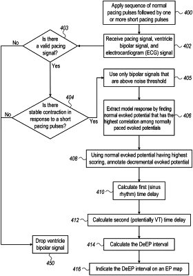
1. A method for evaluation of electrical propagation in a heart of a patient, the method comprising:
applying a pacing signal to the heart of the patient by electrodes on a catheter, the pacing signal comprising (i) a sequence of normal pacing stimuli at sinus-rate intervals, and (ii) one or more abnormal pacing stimuli at abnormal intervals that are shorter than the sinus-rate intervals;
receiving, in response to the applied pacing signal, a cardiac signal that is sensed by the catheter electrodes at a location in the heart;
identifying and annotating a model response from evoked potentials in the cardiac signal caused by the normal pacing stimuli, the annotated model response being one of the evoked potentials having a high correlation of stimulated points of cardiac activity to the other evoked potentials in the cardiac signal;
determining, for each interval between paces of the paced signal, scorings of correlations with the model response along each of the respective intervals between paces of the paced signal;
annotating a first set of the evoked potentials in the cardiac signal caused by the normal pacing stimuli and a second set of the evoked potentials in the cardiac signal caused by the one or more abnormal pacing stimuli, the first and second sets of evoked potentials being other than the evoked potential of the model and being annotated based upon the scorings of correlations, each annotation in each interval of the paced signal being identified as an index that received the highest scoring with the model response for that interval;
determining a first time delay between the normal pacing stimuli and the resulting evoked potentials in the cardiac signal and a second time delay between the abnormal pacing stimuli and the resulting evoked potentials in the cardiac signal;
determining a decremental time delay, the decremental time delay being a time difference between the first time delay and the second time delay; and
presenting an EP map of at least a portion of the heart to a user, with a graphical indication of the decremental time delay presented at the one or more locations in the heart.
|