| CPC A61B 5/055 (2013.01) [A61B 5/0042 (2013.01); G01R 33/50 (2013.01); G01R 33/5608 (2013.01); G01R 33/56308 (2013.01)] | 12 Claims |
|
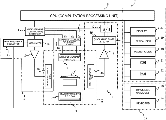
1. A magnetic resonance imaging apparatus comprising:
a static magnetic field generation unit that applies a static magnetic field to an imaging space in which an object under examination is placed;
a gradient magnetic field generation unit that applies a gradient magnetic field to the imaging space;
a transmitting coil that transmits a high-frequency magnetic field to the object under examination in the imaging space;
a receiving coil that receives a nuclear magnetic resonance signal from the object under examination;
a sequencer that performs an imaging sequence by controlling the gradient magnetic field generation unit, the transmitting coil, and the receiving coil to capture an image; and
a computation processing unit coupled to a memory,
wherein the sequencer captures, with respect to an equal imaging region of the object under examination, a morphology image with a morphology expressed therein and a function image with a function expressed therein, and
wherein the memory stores instructions that when executed by the computation processing unit causes the computation processing unit to:
execute processing for deforming the morphology image using a deformation parameter to move positions of one or more structural objects included in the morphology image to respective positions of structural objects of a previously determined standard morphology thereby causing the morphology image to coincide with the standard morphology by performing normalization using non-linear conversion using at least the deformation parameter, and
then to execute processing for deforming the function image using a value of the deformation parameter used in deforming the morphology image to cause a position of a region included in the function image to coincide with a position of a corresponding region of the standard morphology or processing for deforming the standard morphology in an opposite direction using the value of the deformation parameter to cause a position of a region of the structural object of the standard morphology to coincide with a position of a corresponding region included in the function image,
wherein the imaging sequence is a sequence which implements a post labeling delay (PLD) according to a single PLD arterial spin labeling (ASL) method of setting one type of time required from start of labeling of blood to execution of imaging and performing imaging using the one type of time, and
wherein the function image is an image of cerebral blood flow (CBF).
|