| CPC G06T 7/0012 (2013.01) [G06T 7/11 (2017.01); G06T 7/30 (2017.01); G16H 70/60 (2018.01); G06T 2207/10101 (2013.01); G06T 2207/20081 (2013.01); G06T 2207/20084 (2013.01); G06T 2207/30041 (2013.01)] | 17 Claims |

|
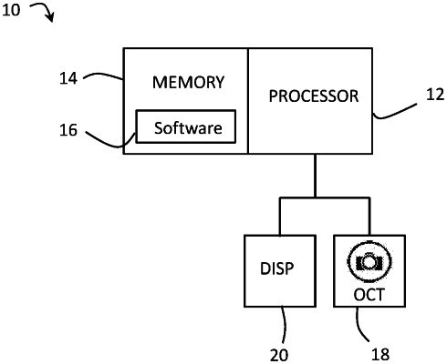
1. A method of analysing images of a retina captured by an Optical Coherence Tomography (OCT) scanner, the method using a processor configured to implement the steps of:
receiving an image of a retina of a patient from an OCT scanner, the image having a plurality of pixels;
segmenting boundaries between layers of the retina for each of the pixels;
determining the layers of the retina using the segmented boundaries;
segmenting regions of pathology of the retina for each of the pixels;
determining a location of the regions of pathology with respect to the determined layers of the retina using the segmented regions;
determining the regions of pathology using the segmented regions and the determined location of the regions;
determining a thickness of the layers of the retina using the segmented boundaries in a neural network model;
determining the thickness of the layers of the retina using a distribution of layer thicknesses derived from known layer thicknesses of a population;
determining a property of the regions of pathology of the retina using the segmented regions;
analysing results of determinations of the regions of pathology and the property of the regions to derive an assessment of the retina of the patient,
wherein the property of the regions of pathology includes an indication of the volume of the regions of pathology; and
outputting the assessment of the retina of the patient.
|