| CPC G01N 33/533 (2013.01) [G01N 21/6428 (2013.01); G01N 33/52 (2013.01); G01N 2021/6439 (2013.01)] | 8 Claims |
|
1. An extracellular vesicle capture and detection system comprising:
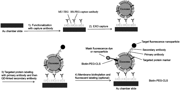
(a) an extracellular vesicle derived from a cancer cell, the extracellular vesicle comprising first and second fluorescent dyes, each having emission spectra that are separated and do not interfere with each other, wherein the first fluorescent dye is a mask labelling agent, and comprises a cholesterol-poly(ethylene) glycol lipophilic linker that binds to an exosome membrane to image and localize the exosome using dark field imaging, and the second fluorescent dye is a target labelling agent and comprises specific binding partners that specifically binds a cancer protein marker, wherein the target labelling agent labels the cancer protein marker to detect, image, and quantify the cancer protein marker on the membrane of the extracellular vesicle, and wherein the extracellular vesicle is bound to a capture molecule covalently bound to a film coating a planar support;
(b) an array comprising a plurality of holes, wherein the array is fixed to the film-coated surface of the planar support to form a plurality of fluid-tight wells;
(c) a laser tuned to emit a wavelength that can effectively excite the two fluorescent dyes;
(d) a signal collection device configured for collecting a signal from the excited fluorescent dyes, and converting the signal into mask and target images, where the fluorescent signals from the first fluorescent dye and the second fluorescent dye are at different wavelengths and wherein the detection system comprises a white light source for dark field imaging and the mask image is an image obtained by dark field imaging; and
(e) software configured to analyze the mask and target images and convert them into a histogram that characterizes the cancer marker present on the exosome.
|