| CPC A61F 9/00781 (2013.01) [A61F 9/0084 (2013.01); A61F 2009/00851 (2013.01); A61F 2009/00872 (2013.01)] | 26 Claims |
|
1. An integrated surgical system for imaging and treating ocular tissue of an eye having a cornea, an iris, an anterior chamber, an irido-corneal angle, and a direction of view, the integrated surgical system comprising:
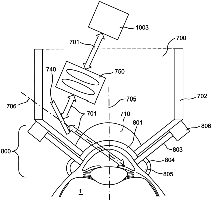
a first optical subsystem configured to establish a common optical path through the cornea and the anterior chamber into the irido-corneal angle, wherein the common optical path is offset from an optical axis within the direction of view;
a laser source configured to output a laser beam;
a second optical subsystem configured to rotate relative to the optical axis, the second optical sub system comprising:
an optical coherence tomography (OCT) imaging apparatus configured to output an OCT beam,
a scanning component optically coupled with the laser source and the OCT imaging apparatus to receive each of the laser beam and the OCT beam, and
focusing optics optically coupled between the scanning component and the first optical subsystem; and
a control system coupled with the laser source and the second optical subsystem and configured to affect operation of the laser source, the OCT imaging apparatus, the scanning component, and the focusing optics to:
obtain a circumferential OCT image of the irido-corneal angle;
obtain an azimuthal OCT image of the irido-corneal angle; and
deliver optical energy through the laser beam in accordance with a treatment pattern for a volume of ocular tissue of the irido-corneal angle, wherein the treatment pattern is based on the circumferential OCT image and the azimuthal OCT image.
|