| CPC A61B 8/523 (2013.01) [A61B 8/085 (2013.01); A61B 8/0883 (2013.01); A61B 8/14 (2013.01); A61B 8/463 (2013.01); A61B 8/483 (2013.01); G01S 15/8993 (2013.01); G06F 16/51 (2019.01); G06F 18/22 (2023.01); G06T 7/0014 (2013.01); G06T 7/10 (2017.01); G06T 7/60 (2013.01); G06T 7/70 (2017.01); G06T 15/005 (2013.01); G06V 10/42 (2022.01); A61B 8/12 (2013.01); G06T 2207/10136 (2013.01); G06T 2207/30048 (2013.01); G06T 2207/30168 (2013.01)] | 20 Claims |
|
1. An ultrasound imaging system comprising:
one or more processors configured to:
receive first volume ultrasound data of a first trans-esophageal scan of an anatomical object within the body obtained by a trans-esophageal echocardiography (TEE) probe positioned at a first position within an esophagus of the body;
receive second volume ultrasound data of a second trans-esophageal scan of the anatomical object obtained by the TEE probe positioned at a second position within the esophagus, wherein the second position is different from the first position;
generate, based on the first volume ultrasound data, a first plurality of two-dimensional (2D) ultrasound images, wherein the first plurality of 2D ultrasound images correspond to a set of pre-defined views of the anatomical object, wherein the first plurality of 2D ultrasound images comprises a first ultrasound image corresponding to a first pre-defined view of the set of pre-defined views;
determine a first quality factor of the first ultrasound image;
generate, based on the second volume ultrasound data, a second plurality of 2D ultrasound images corresponding to the set of pre-defined views, wherein the second plurality of 2D ultrasound images comprises a second ultrasound image corresponding to the first pre-defined view;
determine a second quality factor for the second ultrasound image;
perform a comparison between the first quality factor and the second quality factor:
select, for the first pre-defined view, the first ultrasound image and not the second ultrasound image based on the comparison; and
output to a display in communication with the one or more processors:
a third plurality of 2D ultrasound images corresponding to the set of pre-defined views, wherein the third plurality comprises the first ultrasound image and not the second ultrasound image; and
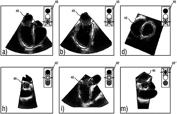
a graphical representation of the first quality factor comprising an icon selected from a plurality of icons, wherein the plurality of icons comprises:
a first icon representative of the first quality factor being within a first range of values; or
a second icon representative of the first quality factor being within a second range of values.
|