| CPC A61B 8/0841 (2013.01) [A61B 8/4209 (2013.01); A61B 8/481 (2013.01); A61J 15/0003 (2013.01); A61J 15/0088 (2015.05); A61M 25/0158 (2013.01); A61B 1/00158 (2013.01); A61B 1/01 (2013.01); A61B 2017/00876 (2013.01); A61B 2017/3413 (2013.01); A61B 2090/378 (2016.02); A61M 2025/0166 (2013.01); A61M 25/10 (2013.01); A61M 2025/1079 (2013.01)] | 28 Claims |

|
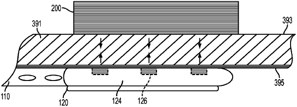
1. A system for coaptive ultrasound visualization, comprising:
a first component configured to be placed on a first side of a plane including at least a portion of a tissue layer, the first component including a magnetic field source;
a second component, having one or more echogenic properties, configured to be placed on a second side of the plane and within an organ, the second component having a proximal end and a distal end, the distal end including a magnetic force receiver configured to interact with a magnetic field generated by the magnetic field source and to generate a force between the first component and the distal end of the second component, wherein the force causes (i) the distal end of the second component to move in a direction within the organ toward the first component and perpendicular to the plane, thereby pushing against a part of the organ and causing an area of coaptation between the part of the organ and the tissue layer while the first component is placed on the first side of the plane and generating the magnetic field and the distal end of the second component is within the organ; and (ii) coordinated movement between the first component and the second component; and
an ultrasound visualization component including an ultrasound probe and a display, the ultrasound probe configured to be placed on the first side of the plane and adjacent the first component, the ultrasound probe configured to generate one or more ultrasonic images of the area of coaptation taken along a line transverse to the plane while the ultrasound probe is adjacent the first component for delivery to the display, and the display configured to output the one or more ultrasonic images of the area of coaptation.
|