| CPC A61B 10/0266 (2013.01) [A61B 10/0096 (2013.01); G06T 7/10 (2017.01)] | 20 Claims |
|
1. A biopsy tissue handling apparatus, comprising:
a tissue holder assembly comprising a tray defining a tissue storage compartment;
a tube defining a vacuum lumen in communication with the tissue storage compartment, the tube being configured to receive a severed tissue specimen and deliver the severed tissue specimen with a fluid through the vacuum lumen so that the severed tissue specimen and the fluid are deposited into the tissue storage compartment;
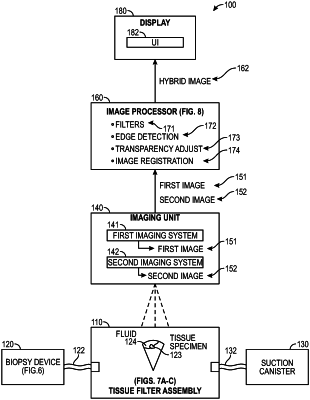
an x-ray imaging system arranged relative to the tissue storage compartment to acquire an x-ray image of the severed tissue specimen and the fluid in the tissue storage compartment, the x-ray image depicting internal structures of the severed tissue specimen and external or surface portions of the severed tissue specimen, wherein the x-ray image includes an attenuated edge of the severed tissue specimen covered by the fluid;
an optical camera imaging system arranged relative to the tissue storage compartment to acquire an optical image of the severed tissue specimen and the fluid in the tissue storage compartment, the optical image depicting only the external or surface portions of the severed tissue specimen, wherein the optical image includes an edge of the severed tissue specimen;
an image processor in communication with the x-ray imaging system and the optical camera imaging system, the image processor being configured to receive the x-ray image and the optical image, detect the edge of the severed tissue specimen depicted in the optical image, and generate a hybrid image based at least in part upon the x-ray image, the optical image, and the detected edge of the severed tissue specimen depicted in the optical image, wherein a transparency of a portion of the optical image corresponding to the detected edge compensates for fluid attenuation of the attenuated edge of the severed tissue specimen depicted in the x-ray image during x-ray image acquisition; and
a display in communication with the image processor, the hybrid image being presented to a user of the biopsy tissue handling apparatus through the display.
|