| CPC G01T 1/2985 (2013.01) [A61B 6/037 (2013.01); A61B 6/4266 (2013.01); A61B 6/4275 (2013.01); A61B 6/4435 (2013.01); G01T 1/20181 (2020.05); G01T 1/20182 (2020.05)] | 15 Claims |

|
1. A positron emission tomography (PET) imaging system, comprising:
a gantry having a patient-receiving tunnel;
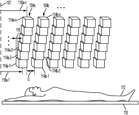
a first detector unit and a second detector unit housed inside the gantry, wherein the first detector unit comprises a plurality of detector elements in a helical arrangement around an axial axis of the PET imaging system, and wherein the second detector unit comprises a plurality of detector elements in a helical arrangement around the axial axis of the PET imaging system,
wherein each of the detector elements in the second detector unit is spaced apart from a corresponding detector element in the first detector unit along a direction parallel to the axial axis of the PET imaging system by an axial gap;
wherein each detector element in the first and second detector units has an axial position measured parallel to the axial axis from an entrance of the patient-receiving tunnel to a geometric center of the detector element, and
wherein each of the first and second detector units comprising its plurality of detector elements arranged so that a set of the plurality of detector elements is positioned such that each detector element in the set is offset from an adjacent detector element in the detector unit in a direction parallel to the axial axis of the PET imaging system such that a maximum difference between axial positions of detector elements in each detector unit is less than or equal to the axial gap.
|