| CPC G16H 20/40 (2018.01) [A61B 18/00 (2013.01); A61B 34/25 (2016.02); A61B 90/37 (2016.02); G16H 30/40 (2018.01); G16H 50/30 (2018.01); G16H 50/50 (2018.01); A61B 2018/00577 (2013.01); A61B 2018/00839 (2013.01); A61B 2018/00904 (2013.01); A61B 2090/364 (2016.02)] | 19 Claims |

|
1. A method, comprising:
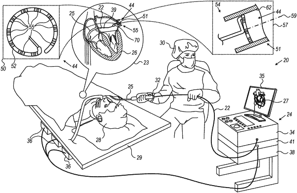
receiving: (i) three-dimensional (3D) data indicative of a selected 3D section that has been ablated in a patient organ in accordance with a specified contour, the selected 3D section comprising an inner wall of a cylindrically-shaped vessel, and (ii) a dataset, which is indicative of a set of lesions formed during ablation of the selected 3D section;
transforming the selected 3D data into a two-dimensional (2D) map by generating a flattened representation of the selected 3D section; and
checking, on the 2D map, whether the set of lesions covers the specified contour.
|