| CPC G01R 33/4828 (2013.01) [A61B 5/055 (2013.01); G06T 7/0012 (2013.01); G06N 3/08 (2013.01); G06T 2207/10088 (2013.01); G06T 2207/20081 (2013.01)] | 18 Claims |

|
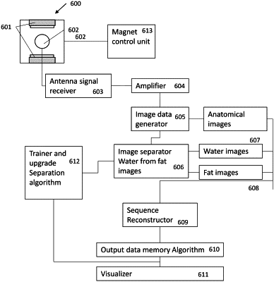
1. A method for separate acquisition and output of diagnostic images in nuclear magnetic resonance imaging based on signals from water and fat, which method comprises:
a) defining at least a slice of an image that passes through a body, or an area of a body, under examination with a pre-established relative orientation;
b) acquiring for at least said slice, or for at least a part of all said slices when there are more than one slice, at least an anatomical image in nuclear magnetic resonance along said slice;
(c) performing, for at least said slice or for at least part of all of said slices when there are more than one, and for each pixel or voxel of the nuclear magnetic resonance image acquired along said slice, a separation of signal components corresponding to an intensity of said pixel or voxel resulting from water within the body under examination from those signal components resulting from fat tissues in said body under examination;
(d) generating for at least said slice, or for at least part of all of said slices when there are more than one, an image called water-only which is generated on the basis of signal components of MRI signals resulting from water-only separated in step (c);
(e) generating for at least a slice, or for at least part of all of said slices, when there are more than one, an image called fat-only which is generated on the basis of signal components of the MRI signals resulting from fat-only separated in step (c);
(f) sorting images related to any single slice in a sequence that for each slice provides firstly for said anatomical image(s) and afterward said water-only image(s) followed by said fat-only image(s);
g) sorting images related to any single slice according by position order of said slices when there are more than one, and wherein
h) the separation of signal components corresponding to intensity of said pixel or voxel resulting from water within the body under examination from those signal components resulting from fat tissues in said body under examination of step c) being performed by a machine learning algorithm that uses automatic learning to configure the machine learning algorithm process based on feedback mechanisms involving training and testing processes with image data; and
using a parameter analysis operator to reduce a number of image pixels, the parameter analysis operator configured to determine an appearance of pixels where an operator identifies regions of the image to be analyzed, wherein parameters of pixel appearance are related to signals resulting from water or fat, submitting to an elaboration process by means of the machine learning algorithm only pixels that fall within the range of said parameters of pixel appearance that are functional to determining the image as resulting from water or fat.
|