| CPC G06N 3/08 (2013.01) [G06F 18/2178 (2023.01); G06F 18/2193 (2023.01); G06F 18/241 (2023.01); G06N 3/04 (2013.01); G06T 7/0012 (2013.01); G06V 10/454 (2022.01); G06V 10/764 (2022.01); G06V 10/82 (2022.01); G06V 20/698 (2022.01); G16H 10/40 (2018.01); G16H 30/20 (2018.01); G16H 30/40 (2018.01); G16H 50/20 (2018.01); G06T 2207/10056 (2013.01); G06T 2207/20081 (2013.01); G06T 2207/20084 (2013.01); G06T 2207/30024 (2013.01); G06T 2207/30096 (2013.01); G06V 2201/03 (2022.01)] | 14 Claims |

|
1. A method to determine a degree of abnormality, the method comprising the following steps:
a) receiving a whole slide image (11, w, 722), the whole slide image (11, w, 722) depicting at least a portion of a cell, in particular a human cell;
b) classifying at least one image tile (13, 601, 721, 721′, 721″) of the whole slide image (11, w, 722) using a neural network (600) to determine a local abnormality degree value (15, a_j, 519, 719, 719′, 719″) associated with the at least one image tile (13, 601, 721, 721′, 721″), the local abnormality degree value (15, a_j, 519, 719, 719′, 719″) indicating a likelihood that the associated at least one image tile depicts at least a part of a cancerous cell; and
c) determining a degree of abnormality (17) for the whole slide image (11, w, 722) based on the local abnormality degree value (15, a_j, 519, 719, 719′, 719″) for the at least one image tile (13, 601, 721, 721′, 721″),
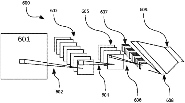
wherein the neural network (600) comprises:
at least fifty layers,
at least twenty pooling layers,
at least forty convolutional layers,
at least twenty kernels in each convolutional layer, and/or
at least one fully connected layer (608).
|