| CPC A61B 6/4405 (2013.01) [A61B 6/4417 (2013.01); A61B 6/4441 (2013.01); H01J 35/147 (2019.05); A61B 6/022 (2013.01); A61B 6/025 (2013.01); H01J 35/18 (2013.01)] | 16 Claims |

|
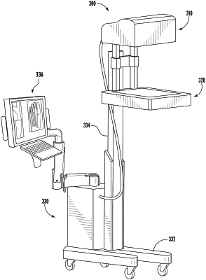
1. A compact x-ray imaging system, comprising:
an x-ray source array comprising a plurality of spatially distributed x-ray focal spots and a digital area x-ray detector;
a collimation assembly connected to an exit window of the x-ray source array configured to substantially collimate x-ray radiation generated from each of the plurality of spatially distributed x-ray focal spots to a surface of the digital area x-ray detector;
an electronic switching device comprising:
a high voltage power supply;
a current source;
a switch configured to sequentially connect the current source to a plurality of field emission cathodes of the compact x-ray imaging system, with a pre-set current value, one at a time, to produce one or more projection images for tomosynthesis reconstruction without any mechanical motion of either the x-ray source array or the digital area x-ray detector; and
a trigger comprising one or more first processors and/or circuitry configured to synchronize detector data collection with x-ray exposure from the plurality of spatially distributed x-ray focal spots; and
a mechanical support configured to enable a position and orientation of the x-ray source array and the digital area x-ray detector to be adjusted such that both upper and lower extremities of a patient can be imaged using tomosynthesis in either a non-load bearing position or a load bearing position;
wherein the compact x-ray imaging system is configured to operate in a plurality of imaging modes;
wherein, when the compact x-ray imaging system is operated in the tomosynthesis imaging mode, a scanning x-ray beam is produced by sequentially activating x-ray beams from the plurality of spatially distributed x-ray focal spots electronically without moving any of the x-ray source array, the digital area x-ray detector, or the patient, in order to collect one or more required projection images for tomosynthesis reconstruction; and
wherein, when the compact x-ray imaging system is operated in the pulsed fluoroscopy imaging mode, x-ray radiation generated from a central focal spot of the plurality of spatially distributed x-ray focal spots is pulsed from about 5 to 30 pulses per second and, for each x-ray pulse, an image of an object being scanned is formed and displayed to produce an x-ray movie of the object.
|