| CPC A61B 5/004 (2013.01) [A61B 5/055 (2013.01); A61B 5/7292 (2013.01); G01R 33/4838 (2013.01); G01R 33/5608 (2013.01); G01R 33/5635 (2013.01)] | 15 Claims |

|
1. A method for imaging a subject, the method comprising:
applying a radiofrequency pulse to the subject;
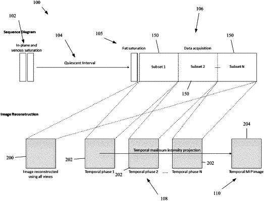
after a quiescent interval, performing a radial acquisition over a duration corresponding to a cardiac cycle of the subject to generate acquisition data;
reconstructing a plurality of 2-D images across a plurality of sequential temporal phases within the duration from the acquisition data, each of the 2-D images depicting a same partial cross-section of the subject and being reconstructed from a continuously sampled subset of the plurality of the sequential temporal phases; and
generating a temporal maximum intensity projection image of the same partial cross-section of the subject by tracking an intensity of each pixel across the plurality of 2-D images and selecting a maximum intensity value for the pixel across the plurality of 2-D images, which corresponds to a maximum intensity during the cardiac cycle, such that vascular stripe artifacts in the temporal maximum intensity projection image are removed relative to the plurality of 2-D images.
|